Degenerative SLAP Tears and Biceps Tendinitis: What the Diagnosis Means After 40
A SLAP tear is an injury to the superior labrum - the rim of cartilage at the top of the shoulder socket, where the biceps tendon anchors. SLAP stands for Superior Labrum Anterior to Posterior, which describes where the tear sits rather than what caused it.
Most patient information about SLAP tears is written about young overhead throwing athletes. But the majority of superior labral tears identified on MRI are degenerative - age-related change in patients over 40 - and these behave quite differently from the tear a twenty-two-year-old pitcher sustains. They arrive differently, they respond to different treatment, and, most importantly, they are far less likely to be the reason the shoulder hurts.
An MRI report that reads “SLAP tear” in a fifty-five-year-old is describing something genuinely present. Whether it is the source of the symptoms is a separate question, and it is the question this article is about.
The Anatomy: Why the Biceps and the Labrum Are One Problem
The labrum is a rim of fibrocartilage around the edge of the glenoid, the shallow socket of the shoulder joint. It deepens the socket and gives the surrounding ligaments something to attach to.
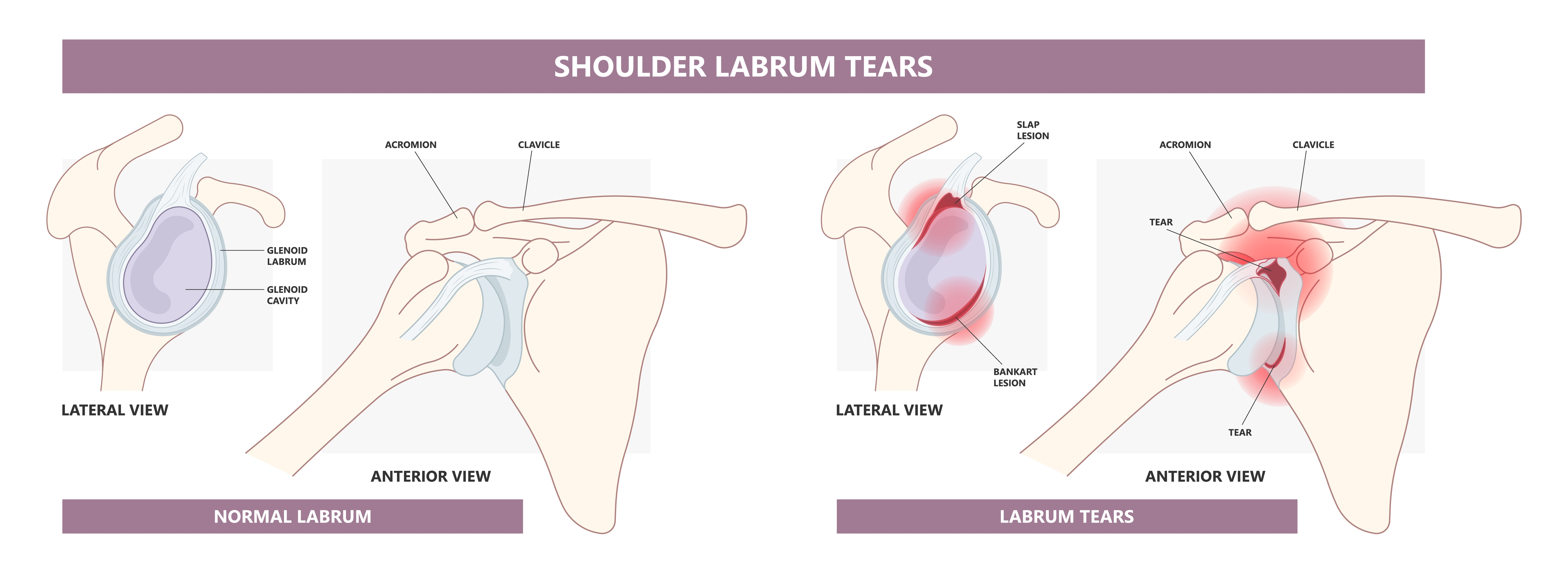
Caption: Where a SLAP tear sits. Left: a normal labrum, the rim of cartilage around the edge of the socket, shown from the side and from the front. Right: the two most common tear locations. A SLAP lesion is at the top of the socket, where the biceps tendon anchors - the subject of this page. A Bankart lesion is at the front rim and occurs when the shoulder dislocates forward, which is a different problem.
The long head of the biceps tendon runs from the muscle in the upper arm, up through a narrow groove at the front of the humerus, into the shoulder joint, and attaches to the top of the glenoid - where it blends directly into the superior labrum. The tendon and the top of the labrum are continuous tissue. There is no clean dividing line between them.
Two consequences follow from that arrangement, and they shape everything below.
The superior labrum and the biceps anchor are functionally a single structure. A tear at that attachment is, mechanically, a problem with the biceps anchor. This is why the operation most often used for these tears addresses the biceps tendon rather than the labrum itself - treating the structure that is generating the symptoms rather than the one the MRI happened to name.
The long head of the biceps is a well-recognized source of shoulder pain in its own right. It travels through a tight bony groove, is subject to repetitive traction and friction with every overhead reach, and is densely innervated. Degeneration and inflammation within it - biceps tendinitis, or more accurately biceps tendinopathy - produce a characteristic pain at the front of the shoulder. In many patients with a labral finding on imaging, the biceps tendon is where the symptoms are actually coming from.
A note on terminology. “Tendinitis” implies inflammation, and acute inflammation is part of the picture in some cases. But in most patients over 40, what is present in the tendon is tendinopathy - degenerative change in the structure of the collagen itself, which weakens the tissue. The distinction matters because it explains why anti-inflammatory treatment alone often produces only temporary relief, and why the tissue does not simply heal back to normal.
Degenerative and Traumatic Tears Are Two Different Situations
| Traumatic SLAP tear | Degenerative SLAP tear | |
|---|---|---|
| Typical patient | Under 35, overhead athlete | Over 40 |
| Mechanism | Throwing, a fall on an outstretched arm, a traction injury | No specific injury - gradual change over years |
| Onset | Often traceable to a single event | Gradual, with no clear starting point |
| Tissue quality | Healthy tissue, torn | Frayed and degenerated |
| Usually found | As an isolated finding | Alongside rotator cuff disease, biceps degeneration, or arthritis |
| Likelihood the finding explains the pain | Reasonably high | Considerably lower - see the section below |
| Operation usually considered | SLAP repair | Biceps tenodesis |
These are the same words on a radiology report describing two clinically different situations. A traumatic tear in healthy tissue in a young athlete is a structural injury to a structure that was previously normal. Degenerative fraying in a fifty-year-old is a change that accumulates in most shoulders over time, in the same way that the rotator cuff, the meniscus in the knee, and the discs in the spine all change with age.
What an MRI Finding of “SLAP Tear” Means After 40
This is the most useful thing on this page, and it is the part most often missing from the conversation.
Superior labral tears are read on MRI in a very large proportion of people who have no shoulder symptoms at all. In a study of middle-aged patients with entirely asymptomatic shoulders, MRI-diagnosed superior labral tears were present in 55 to 72 percent of those aged 45 to 60. Imaging-based studies show the prevalence climbing steadily with age, with age emerging as the single strongest predictor of a superior labral abnormality once other factors are accounted for.
Read that the other way round: in this age group, finding a superior labral tear on an MRI is closer to the expected result than to an abnormal one. These changes are also seen in anatomical studies of shoulders from people who never reported shoulder problems, which is what you would expect of an age-related process rather than an injury.
This does not mean the finding is meaningless, and it does not mean the pain is imagined. It means the finding on its own does not establish where the pain is coming from. The shoulder hurts for a reason, and the job is to identify which structure is responsible - which requires the history, the examination, and often a diagnostic injection, considered alongside the imaging rather than after it.
The same principle applies across the shoulder. Rotator cuff abnormalities are found on imaging in a similarly high proportion of people without symptoms, and for the same reason. A scan shows what a structure looks like. It cannot show what hurts.
Symptoms
Degenerative SLAP tears and biceps tendinopathy typically produce pain at the front of the shoulder, worse with overhead reaching and with lifting away from the body, often with a deep clicking or catching sensation.
- Pain at the front of the shoulder, frequently localized precisely to the biceps groove. Patients often point to the same spot with one finger, which is one of the more useful pieces of information in the whole assessment
- Pain with overhead activity and with lifting the arm away from the body
- Pain with resisted elbow flexion or with turning the forearm palm-up - opening a heavy door, turning a stiff doorknob, carrying a grocery bag palm-up
- Deep clicking, catching, or popping within the shoulder
- Night pain, particularly when lying on that side
- A sense of weakness, which is usually the arm guarding against pain rather than true loss of strength
- Symptoms that overlap with the rotator cuff. Biceps and cuff problems travel together, and separating them is often the central diagnostic question
In younger throwing athletes the presentation is different: loss of velocity or command, deep pain at the back of the shoulder in the late-cocking phase of throwing, and the sensation athletes describe as a “dead arm.” That is a distinct problem, assessed differently, and is not the subject of this page.
How the Diagnosis Is Made
No single physical examination test for a superior labral tear is reliably accurate on its own. O’Brien’s active compression test, Speed’s test, Yergason’s test, and the biceps load tests are all in use, and each has modest accuracy when used in isolation. Used in a structured combination, they perform considerably better - and that combination is what an examination for this problem should look like.
The Three-Pack Examination
The three-pack examination is a set of three tests performed together to assess the biceps-labrum complex, including the part of the biceps tendon that sits outside the joint in the bicipital tunnel - a region that arthroscopy alone does not fully visualize.
- The active compression test (O’Brien’s test). The arm is held forward at shoulder height, brought slightly across the body, and turned thumb-down while downward pressure is resisted. The test is then repeated palm-up. Pain that is worse thumb-down and eases palm-up points toward the biceps-labrum complex
- The throwing test. The arm is placed in the late-cocking position of a throw - out to the side and rotated back - and the patient performs a throwing motion. Pain reproduced at the front of the shoulder is a positive test
- Palpation of the bicipital tunnel. Direct pressure over the biceps tendon in its groove, a few centimeters below the front of the acromion. This is often the test that matches what the patient already told you, because the spot they point to with one finger is frequently this exact place
What this combination is good at is exclusion. Each individual test has high sensitivity but only moderate specificity, meaning a positive result narrows the field without confirming the diagnosis. Taken together, though, the negative predictive value is high - reported at 93 to 96 percent for disease within the bicipital tunnel when tunnel palpation and the O’Brien test are both negative. In plain terms: a negative three-pack is strong evidence that the biceps-labrum complex is not the problem, which redirects the search rather than leaving it open.
That is a genuinely useful property in a diagnosis where imaging over-reads. It also explains the sequence - the examination narrows the possibilities, and a diagnostic injection then tests the leading one directly.
The examination is also doing something broader than testing for one structure. A shoulder examination assesses the rotator cuff, the biceps, the acromioclavicular joint, range of motion, and the neck, because the same front-of-shoulder pain can arise from several of these and they frequently coexist. What the examination establishes is not usually a single diagnosis but a shortlist.
MRI. MRI is the imaging study for this problem. In my practice a standard MRI without contrast is usually sufficient, particularly when it is performed on a 3 Tesla scanner - a higher field strength magnet than the 1.5 Tesla machines still in common use, which produces finer resolution and shows the labrum and biceps anchor well without anything being injected.
MR arthrography - MRI performed after contrast is injected into the joint - outlines the labrum by filling the space around it, and some centers use it routinely. It is not our default. It is most useful in the specific situation where the MRI has not answered the question and clinical suspicion remains high, and reserving it for that situation avoids an injection that in most cases would not change the plan.
Both studies over-read in this age group, for the reasons set out above. An MRI in a fifty-five-year-old will frequently report a superior labral tear, and that report is usually an accurate description of the tissue. What it does not establish is whether that tissue is the source of the symptoms.
MRI is also ordered for reasons other than the labrum: to assess the rotator cuff, to establish what else is present in the joint before any intervention is planned, and to look for the conditions that produce similar symptoms.
A diagnostic injection is frequently the most informative test available. An ultrasound-guided injection of local anesthetic into the biceps sheath tests the question directly: if the pain resolves for the duration of the anesthetic, the biceps is the pain generator. If it does not, the pain is coming from somewhere else, and that is equally useful to know. In a region where imaging over-reads and examination testing under-performs, this is often what settles the question - and it does so before an operation rather than after.
The diagnosis rests on all of it together. History, examination, imaging, and frequently a diagnostic injection. Not on the imaging report alone.
Conditions That Produce Similar Symptoms
Front-of-shoulder pain has several possible sources, and more than one is often present at the same time.
- Rotator cuff disease - particularly subscapularis and anterior supraspinatus involvement, which produces pain in a similar location. Cuff and biceps pathology coexist very commonly
- Acromioclavicular joint arthritis - pain at the top of the shoulder that can be difficult to separate from biceps pain by description alone
- Adhesive capsulitis - in its early painful phase, before stiffness is obvious, frozen shoulder can present as unexplained shoulder pain. This is a distinction that matters, because the sequence of treatment differs entirely
- Scapular dyskinesis - abnormal shoulder blade mechanics increase load on the biceps anchor and are frequently part of the picture rather than a separate diagnosis
- Neck-related pain - cervical spine pathology refers pain to the shoulder region and can coexist with a genuine shoulder problem
This is why the diagnostic injection is so useful. When several plausible sources are present at once, a test that isolates one of them is more useful than a scan that shows all of them.
Non-Surgical Treatment
Non-surgical treatment is the starting point for degenerative superior labral tears and biceps tendinopathy, and it resolves symptoms for a substantial proportion of patients.
Activity modification. Reducing overhead loading and heavy palm-up lifting for a period, rather than indefinitely. The purpose is to lower the load on an irritated tendon enough for it to settle, not to stop using the arm.
Physical therapy targeting the rotator cuff, the scapular stabilizers, and posterior capsular mobility. Tightness at the back of the shoulder shifts load onto the superior labrum and the biceps anchor, and addressing it is often what changes the symptoms. Therapy also addresses the postural mechanics that increase the load in the first place, which matters particularly for desk workers.
For structured home exercise, the AAOS shoulder conditioning program is a reasonable general resource to work alongside a therapy program.
Anti-inflammatory medication, which helps most in the phase where inflammation is a real component. In longstanding degenerative tendinopathy, the benefit is usually more limited.
Corticosteroid injection. An injection into the biceps sheath or the glenohumeral joint serves both purposes at once - it treats the inflammation and it tells you something about where the pain is coming from. Ultrasound guidance matters here, because the biceps sheath is a small target and placing the medication within it concentrates the effect on the tissue in question.
How long to give it. Degenerative tendon problems respond slowly, and a program abandoned after a handful of visits has not really been tested. In my practice I look for a dedicated trial of three months or so before the conversation turns toward surgery, unless something in the presentation argues for moving sooner.
When Surgery Is Considered
Surgery becomes a reasonable discussion when pain persists despite a dedicated trial of non-surgical treatment, and the biceps or the labrum has been reasonably established as the source of symptoms rather than assumed from a scan.
That second condition is the one that does the work. An operation directed at a structure that is not generating the pain will not relieve the pain, however well the operation is performed. This is why the diagnostic injection carries so much weight in this particular diagnosis.
When surgery is appropriate, the choice of operation matters as much as the decision to operate.
The Operations
Biceps Tenodesis
Tenodesis detaches the long head of the biceps from its attachment at the top of the labrum and fixes it to the humerus, outside the joint. The degenerated tendon and the painful anchor are taken out of the joint entirely, and the biceps muscle keeps a solid attachment to bone - which preserves both its strength and the normal contour of the arm.
The logic is different from a repair. A repair attempts to heal degenerated tissue back onto bone. A tenodesis removes the problem tissue from the joint and re-anchors the tendon where it is not under the same stress. For degenerative tears, for patients over 40, and for people who are not overhead throwing athletes, this is generally the operation I recommend.
Where the tendon is fixed, and how. I perform both arthroscopic suprapectoral and open subpectoral tenodesis, and the choice depends on the case.
- Arthroscopic suprapectoral tenodesis fixes the tendon at or just below the top of the bicipital groove and is performed entirely through the scope, at the same sitting as the diagnostic arthroscopy and any other work being done in the joint.
- Open subpectoral tenodesis fixes the tendon lower, below the groove, through a small incision beneath the pectoralis major. Because it moves the fixation point below the groove, it takes the whole length of the groove out of the picture as a potential source of ongoing pain.
The decision rests on what is found at the time of surgery - how much of the groove is involved, the condition of the tendon along its length, what else is being addressed in the same operation, and patient factors. Both are well-established techniques with good results, and the choice is a technical one rather than a difference in what the operation is trying to achieve.
Biceps Tenotomy
Tenotomy releases the tendon from its anchor without reattaching it. The tendon retracts down into the arm and scars in place on its own.
It is a simpler procedure with no post-operative restrictions, and it reliably relieves the pain, because the mechanism is the same - the painful tissue is no longer under tension inside the joint. The trade-offs are a visible “Popeye” deformity in a proportion of patients, where the retracted muscle belly bunches in the lower arm, and some aching or cramping with sustained activity in a smaller proportion.
It suits some patients well - typically older, lower-demand patients for whom avoiding a period of restrictions matters more than the appearance of the arm. The trade-offs should be set out clearly and letting the patient weigh, rather than deciding on their behalf.
SLAP Repair
Repair reattaches the superior labrum to the glenoid with suture anchors. It is the operation designed for the injury pattern the condition was originally described in: a traumatic tear in healthy tissue in a younger patient, most often an overhead athlete.
In degenerative tissue, the calculation is different. Sutures hold in healthy labrum. Frayed, degenerated tissue is less reliable to repair, and repairing the biceps anchor in place keeps the degenerated tendon inside the joint, where it can continue to generate symptoms. Repair in this group is associated with a higher rate of post-operative stiffness and a higher rate of further surgery.
What the Evidence Shows, and What It Does Not
This is an area where the evidence is real but more nuanced than either “repair fails after 40” or “repair works fine.”
On reoperation. A systematic review comparing the two operations found reoperation in 12 percent after SLAP repair and 6 percent after biceps tenodesis. A further systematic review, in patients under 40, reported reoperation ranging from 3 to 16 percent after repair and 0 to 6 percent after tenodesis - around a fourfold difference in the likelihood of needing further surgery. These figures come from studies of younger patients, which requires precision: the reoperation advantage for tenodesis is not something that only appears after 40.
On function and satisfaction. In younger patients, pain scores, functional scores, and satisfaction are broadly similar between the two operations. Return to play is comparable - around 78 percent after tenodesis and 68 percent after repair, a difference that did not reach statistical significance, including within the subgroup of overhead athletes. The case for tenodesis in younger patients rests on the lower reoperation rate rather than on better function.
On age specifically. The picture is genuinely mixed. One five-year follow-up of 107 patients found no significant difference in outcome between patients over and under 40. Other work has found associations between age over 40 and both poorer functional scores and a higher complication rate, and a large series in active military patients reported a 37 percent failure rate and a 28 percent revision rate after repair. The honest summary is that repair in older patients is less predictable rather than uniformly unsuccessful, and that the trend in practice has moved toward tenodesis in this group on the basis of that unpredictability.
On the underlying reasoning. The most persuasive argument for tenodesis in a degenerative tear is not statistical at all. It is that the operation matches the problem: degenerated tissue is unreliable to heal, and removing it from the joint addresses the symptoms rather than attempting to restore an anatomy that has been changing gradually for years.
Recovery After Biceps Tenodesis
Recovery is generally quicker and more predictable than after a SLAP repair, which is part of the reason the operation is preferred in this group.
| Period | What to expect |
|---|---|
| Weeks 0–2 to 4 | Sling for comfort and to protect the fixation. The hand, wrist, and elbow are used for light everyday tasks from the outset. |
| To about 6 weeks | No active elbow flexion against resistance and no lifting palm-up. The restriction protects the fixation while the tendon incorporates into bone. |
| 6–12 weeks | Progressive strengthening, including the biceps. |
| 3–4 months | Return to full activity, including gym work and sport. |
These timelines shift when the tenodesis is done alongside another procedure. If a rotator cuff repair is performed at the same time, the cuff protocol governs the recovery, because it is the more protective of the two - see rotator cuff repair surgery and recovery for what that involves.
Frequently Asked Questions
What is a SLAP tear?
A SLAP tear is an injury to the superior labrum at the top of the shoulder socket, where the biceps tendon attaches. SLAP stands for Superior Labrum Anterior to Posterior, which describes the location of the tear. Tears can be traumatic - from throwing or a fall - or degenerative, developing gradually with age.
My MRI says I have a SLAP tear. Is that why my shoulder hurts?
Not necessarily. Superior labral tears are found on MRI in 55 to 72 percent of people aged 45 to 60 who have no shoulder symptoms at all, and the rate rises with age. The finding is usually a real description of the tissue, but on its own it does not establish where the pain is coming from. That takes an examination and often a diagnostic injection alongside the scan.
Does a SLAP tear always need surgery?
No. Most degenerative superior labral tears respond to activity modification, physical therapy, and injections. Surgery is considered when pain persists despite a dedicated trial of non-surgical treatment and the biceps or labrum has been established as the source of symptoms.
Why is biceps tenodesis usually preferred over SLAP repair after 40?
Because degenerated tissue does not heal reliably, and repairing the anchor in place leaves the degenerated tendon inside the joint. Tenodesis takes the painful tissue out of the joint and re-anchors the tendon to bone below it. Repair in older patients is associated with more post-operative stiffness and a higher rate of further surgery, and recovery after tenodesis is faster and more predictable.
What is the difference between suprapectoral and subpectoral tenodesis?
They differ in where the tendon is fixed to the bone. Suprapectoral tenodesis fixes it at the top of the bicipital groove and is done arthroscopically. Subpectoral tenodesis fixes it below the groove through a small incision, which removes the whole groove as a possible source of ongoing pain. Both are well-established, and the choice depends on what is found at surgery and what else is being treated at the same time.
What is the “Popeye” deformity?
It is a visible bulge in the lower part of the upper arm that occurs when a released biceps tendon retracts. It happens in a proportion of patients after biceps tenotomy and is uncommon after tenodesis, where the tendon is reattached to bone. It is a cosmetic change and does not meaningfully affect strength for most daily activities, but it matters to some patients and should be discussed before surgery rather than after.
Will I lose arm strength after biceps tenodesis?
Most patients notice no functional difference. The long head of the biceps contributes less to elbow flexion and forearm supination strength than people expect, and tenodesis preserves the tendon’s attachment to bone. Measurable differences in laboratory testing are small and rarely significant in daily life.
Can biceps tendinitis heal on its own?
Mild, recent cases often settle with activity modification, physical therapy, and time. Longstanding degenerative change within the tendon is less likely to resolve completely, though symptoms can be well controlled without surgery in many patients.
How long is recovery after biceps tenodesis?
Typically a sling for two to four weeks, no resisted elbow flexion or palm-up lifting for about six weeks, progressive strengthening from six to twelve weeks, and return to full activity at around three to four months. Recovery is generally faster and more predictable than after a SLAP repair.
Do I need an MRI with dye injected into the joint to diagnose a SLAP tear?
Usually not. A standard MRI is generally sufficient, particularly on a 3 Tesla scanner, which produces higher-resolution images than the more common 1.5 Tesla machines. An MR arthrogram - where contrast is injected into the joint first - is most useful when the standard MRI has not answered the question and there is still a strong clinical suspicion.
Is a SLAP tear the same as a rotator cuff tear?
No. They involve different structures - the labrum and biceps anchor in one case, the rotator cuff tendons in the other. They frequently occur together in the same shoulder, and separating which one is producing the symptoms is often the main diagnostic question.
Front-of-shoulder pain that will not settle, or an MRI report you would like a second read on?
Dr. Gabriella Ode is a fellowship-trained shoulder and sports medicine surgeon at the HSS Sports Medicine Institute, with offices in Manhattan and Brooklyn. Request an appointment or request a remote second opinion.
Financial disclosure.
Dr. Ode is a paid consultant for Advita Ortho and ConMed, manufacturers of orthopaedic implants and surgical instrumentation. These relationships are reported publicly through the CMS Open Payments database. She receives no compensation for this website, and no compensation is tied to any individual patient’s treatment decision. Her complete financial disclosures are available here.
This article is for general education and is not a substitute for individual medical advice. Please consult a physician about your specific condition.