Degenerative Rotator Cuff Tears: Why They Happen and How They’re Diagnosed
A degenerative rotator cuff tear is a tear that develops gradually from age-related tendon wear rather than from a single injury. It is far more common than most people realize. When researchers performed MRI scans on a random sample of the general population aged 41 to 76 - not patients, just people - nearly 99% had some rotator cuff abnormality, and 96% of shoulders with no symptoms at all had one.
That last point is the reason this article exists. The single most common source of confusion after an MRI report is the assumption that a tear seen on imaging must be repaired. Often it should not be - and sometimes the tear isn’t even what’s causing the pain.
This page covers what a degenerative tear is, why it happens, and how it’s diagnosed. Treatment is covered separately:
- Non-surgical treatment of rotator cuff tears - what a proper program involves, how long to give it, and what the evidence shows
- Rotator cuff repair surgery - when it’s warranted, what the operation involves, and recovery
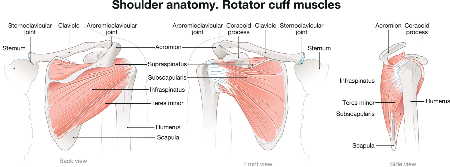
Caption: The rotator cuff is four muscles and their tendons, wrapping the top of the arm bone like a cuff around a wrist. From left: the back view, the front view, and the side view of the same shoulder. The supraspinatus runs across the top and is the tendon most commonly torn. The infraspinatus and teres minor sit behind it and rotate the arm outward. The subscapularis lies at the front and rotates it inward. Together they hold the ball of the shoulder centered in its socket while the larger muscles move the arm — which is why a problem here affects almost everything the shoulder does.
A note on the anatomy, because it explains the rest of this page. The four tendons do not simply move the arm - their main job is to keep the ball of the joint centered in the socket while the much larger deltoid does the lifting. That is why the cuff can be torn and still function reasonably well when the remaining tendons compensate, and also why a large tear can leave someone unable to lift the arm at all despite a perfectly strong deltoid.
Degenerative vs. Traumatic Tears
A degenerative tear develops slowly as tendon tissue deteriorates with age. A traumatic tear happens suddenly, from a specific event such as a fall or a heavy lift.
The distinction drives nearly every treatment decision.
| Degenerative | Traumatic | |
|---|---|---|
| Onset | Gradual, often years | Sudden, identifiable moment |
| Typical age | Over 50 | Any age |
| Tissue quality | Poor - thinned, degenerated | Usually healthy |
| Healing potential after repair | Lower | Higher |
| Urgency | Low; time to try non-surgical care | Higher; early repair often preferred |
| Non-surgical success | Frequently good | Lower for large acute tears |
Why urgency differs. A healthy tendon torn acutely in a younger patient retracts and its muscle begins to atrophy, so timely repair matters - evidence suggests earlier repair is associated with lower retear rates and better functional outcomes. A degenerative tear in a 68-year-old has usually been present for years already, so there is time to try physical therapy first.
Why Degenerative Tears Happen
The way I explain this to patients: a degenerative tear is wear, not injury.
Think about a well-worn pair of jeans. They don’t tear because of something you did on one particular day. The fabric thins gradually at the spot that takes the most stress - the knee, the seat - and eventually it gives way. Nothing dramatic happened. It simply wore out where it was always going to wear out.
Or think about a carpeted room. The patch of rug that wears through first is the one along the path everyone walks. That isn’t bad luck. It’s the area that took the most traffic.
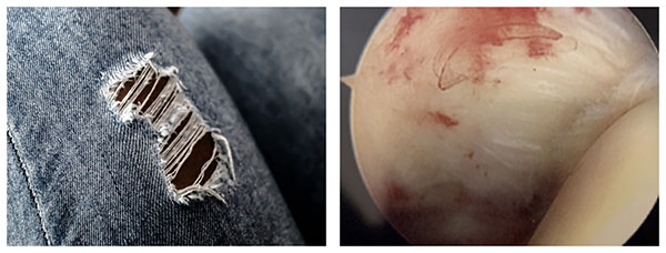
Caption: The same process, in two materials. Left: a worn pair of jeans. The fabric has thinned across a wide area, and within the worn patch some threads have failed while others still span the gap. Right: the view through an arthroscope from inside my patient’s shoulder joint, looking up at the undersurface of the rotator cuff tendon. This is a low-grade partial-thickness tear — the tendon surface has become roughened and irregular where it has worn, but the tendon is still attached. Neither of these was caused by a single event.
The rotator cuff behaves the same way. The tendon wears where it is loaded most, and that location is predictable - which is why the supraspinatus, the tendon at the top of the shoulder, is the one most commonly torn. Decades of reaching, lifting, and carrying concentrate stress in the same small area.
This also explains something patients often find confusing: partial-thickness tears. Jeans go thin before they go through. So do tendons. A partial-thickness tear is the thin spot - the tendon is worn but not yet torn all the way across. It’s the same process caught at an earlier stage, and it’s why a tear can enlarge over time without any new injury.
Compare the two images above, because they are showing you the same thing. On the left, the denim has thinned across a broad area and only then given way in the middle, with individual threads still bridging the gap. On the right is the real equivalent inside a shoulder - an arthroscopic view looking up at the undersurface of the rotator cuff tendon, where the surface has become roughened and irregular from wear. That roughened area is the partial-thickness tear. There is no clean edge, no moment of injury, and the tendon is still attached. It has simply worn thin where it was always going to wear thin.
Articular-sided is the term you may see on your MRI report for this pattern - it means the wear is on the joint side of the tendon, the surface facing into the shoulder, which is the side visible in that arthroscopic image. It is the most common location for a degenerative partial tear, and it’s often why a tear is present without ever having been felt as an injury.
It also explains why “what did I do to cause this?” usually has no satisfying answer. Most of the time, nothing. That’s how wear works.
The specific factors behind that wear:
Blood supply. The supraspinatus tendon - the most commonly torn - has a relatively poor blood supply near its attachment. Less blood means less capacity to repair the microdamage of daily use.
Cumulative loading. Decades of overhead activity produce microtrauma that gradually outpaces healing.
Age-related tendon changes. Collagen organization degrades and tensile strength declines with age. This is normal biology, not a disease.
Anatomy. Acromion shape affects the space the tendon passes through, though the older “impingement causes tears” model is now understood to be an oversimplification.
Systemic factors. Smoking, diabetes, high cholesterol, and thyroid disease are all associated with tendon degeneration and with poorer healing after repair. Smoking is the modifiable factor with the largest effect on repair healing - and I say so plainly to anyone considering surgery.
Genetics. Family clustering of rotator cuff tears is well documented.
Symptoms
The classic pattern is pain over the outer upper arm, worse at night and worse with reaching overhead, along with weakness that has developed gradually.
- Pain location: the outside of the upper arm, often to about the deltoid insertion. Notably not the top of the shoulder (suggests the AC joint) and not radiating past the elbow with numbness (suggests the neck)
- Night pain, especially lying on that side - one of the most consistent features and often the reason people seek care
- Weakness lifting or holding the arm out, sometimes noticed only as difficulty with a specific task
- Painful arc between roughly 60 and 120 degrees of elevation
- Loss of motion - usually mild; profound stiffness suggests frozen shoulder instead
- What you generally won’t have: grinding, clicking, or a catching sensation. Those are mechanical symptoms, and they point toward arthritis in the joint or a labral problem rather than a rotator cuff tear
Important: many tears cause no symptoms whatsoever. A tear found incidentally on an MRI ordered for another reason does not by itself require treatment.
How Common Is This, Really?
Far more common than most people - including many physicians - assume.
In 2026, a Finnish population study published in JAMA Internal Medicine did something that had never been done properly before: rather than scanning patients who came in with shoulder pain, the investigators took a nationally representative random sample of 602 adults aged 41 to 76 from the general population and performed high-resolution MRI on both shoulders, whether or not those shoulders hurt.
Nearly 99% had at least one rotator cuff abnormality. Broken down: 25% had tendinopathy, 62% had a partial-thickness tear, and 11% had a full-thickness tear.
The finding that matters most: abnormalities were present in 96% of shoulders that had no symptoms at all - compared with 98% of shoulders that did hurt. Full-thickness tears were somewhat more common in painful shoulders (14.6% vs. 6.5%), but once the researchers accounted for age, other findings on the scan, and the physical examination, even that difference largely disappeared.
Age drove the pattern, as you’d expect from wear. No full-thickness tears at all were found under age 45. By ages 45 to 49 the figure was 4%. By age 70 and over, it was 28%.
What I take from this, and what I want you to take from it:
A tear on your MRI is not, by itself, an explanation for your pain. Something close to a coin flip of your uncomplaining, symptom-free peers has one too. Finding a tear tells us it’s there; it does not establish that it’s the source of your problem.
Your tear is very unlikely to be something you did wrong. At these prevalence rates, cuff wear is closer to gray hair than to injury - a near-universal consequence of using a shoulder for several decades.
The examination and your history do the diagnostic work; the scan characterizes what we find. This is precisely why I don’t treat scans. I treat patients, and the MRI is one input among several - most useful for defining what a tear looks like once the clinical picture already points to the cuff, and for planning if surgery is on the table.
(Ibounig T, Järvinen TLN, Raatikainen S, et al. Incidental Rotator Cuff Abnormalities on Magnetic Resonance Imaging. JAMA Intern Med. 2026;186(4):406–414.)
Diagnosis
Diagnosis begins with a physical examination. Specific strength tests isolate the individual cuff tendons - the empty can test for supraspinatus, external rotation strength and lag signs for infraspinatus and teres minor, belly-press and lift-off for subscapularis. Examination also assesses passive motion, scapular mechanics, and neck contribution.
X-rays don’t show tendons but reveal a great deal: arthritis, calcific deposits, acromion shape, and - critically - the acromiohumeral interval. Narrowing there suggests a large chronic tear and possible cuff tear arthropathy.
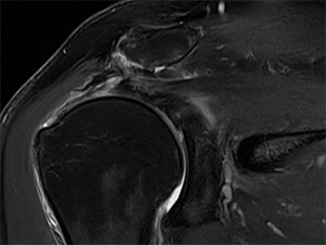
Caption: MRI of a full-thickness rotator cuff tear. The bright white signal is fluid sitting in the gap where the supraspinatus tendon should be attached, and the torn end of the tendon has pulled back to the middle of the humeral head.
MRI is the definitive study. It defines whether a tear is partial or full-thickness, its size, how far the tendon has retracted, and the condition of the muscle.
Retraction matters, because it changes what’s possible. A torn tendon behaves like an elastic band that has been cut - it pulls back toward the muscle. In the image above, the tendon has retracted as far as the middle of the humeral head. The further a tendon has withdrawn, and the longer it has sat there, the harder it is to bring back to bone without tension, and tension is what makes a repair fail.
Muscle quality is often more important than tear size. A tendon whose muscle has undergone significant fatty infiltration and atrophy will not regain function even after a technically perfect repair - a finding that frequently, and appropriately, redirects the whole conversation.
Ultrasound is a reasonable alternative in experienced hands: dynamic, inexpensive, and no tube.
CT arthrogram is used occasionally, and the option exists if you need it. Contrast dye is injected into the joint and a CT scan is then performed; a tear shows up as dye leaking through the gap in the tendon. I reserve it for two situations - when a patient can’t have an MRI, which most often means certain pacemakers or other implanted devices, and when ultrasound either isn’t available or hasn’t given a clear answer.
If you’ve been told you can’t have an MRI, that does not leave you without a way to get a diagnosis. Raise it at your visit rather than assuming the question can’t be answered.
What Happens Next
Having a tear on an MRI is the beginning of a conversation, not the end of one. The two questions that follow are covered in detail on their own pages:
→ Non-surgical treatment of rotator cuff tears What a proper program actually involves, how long to give it before deciding, and what the ten-year evidence shows about patients who never had surgery.
→ Rotator cuff repair surgery When repair is genuinely warranted and when it isn’t, what the operation involves, the sling protocol, and the full recovery timeline.
Frequently Asked Questions
Can a rotator cuff tear heal on its own?
A full-thickness tear will not heal back to bone without surgery. However, symptoms frequently resolve without repair - many patients become pain-free and functional through physical therapy despite a persistent tear, because the surrounding muscles compensate effectively.
What is the difference between a partial-thickness and a full-thickness rotator cuff tear?
A partial-thickness tear is a worn, thinned area where some tendon fibers have failed but others are still attached. A full-thickness tear goes all the way through, leaving a gap where the tendon should meet the bone. Think of jeans: they go thin at the knee before they go through. Partial tears are the thin spot, and they can enlarge over time without any new injury.
What does “articular-sided” partial-thickness tear mean?
It means the wear is on the joint side of the tendon - the surface facing into the shoulder joint - rather than on the outer, bursal side. It’s the most common location for a degenerative partial tear. Seen through an arthroscope, a low-grade articular-sided tear looks like a roughened, irregular patch on an otherwise smooth tendon surface: worn thin, but still attached.
How common are rotator cuff tears?
Much more common than most people expect. A 2026 population study in JAMA Internal Medicine performed MRI scans on both shoulders of 602 randomly selected adults aged 41 to 76 - not patients, just members of the general public. Nearly 99% had at least one rotator cuff abnormality: 25% tendinopathy, 62% a partial-thickness tear, and 11% a full-thickness tear. Full-thickness tears rose from none under age 45 to 28% at age 70 and above.
Can you have a rotator cuff tear with no pain?
Yes, and it’s common. In that same study, rotator cuff abnormalities were present in 96% of shoulders that had no symptoms at all. Full-thickness tears were found in 6.5% of pain-free shoulders. This is why a tear on an MRI does not by itself explain shoulder pain or establish that surgery is needed - the physical examination and your history do most of the diagnostic work.
Did I cause my rotator cuff tear?
Almost certainly not. Degenerative tears come from the accumulated wear of using a shoulder for several decades, not from anything you did wrong. Given that nearly all adults over 40 show some rotator cuff wear on MRI, this is closer to a normal age-related change than an injury.
How do I know if my rotator cuff tear is getting worse?
Warning signs include increasing weakness, new difficulty lifting the arm overhead, and progressively worse night pain. Some degenerative tears enlarge over time; many remain stable for years. Declining function calls for prompt evaluation - progression to cuff tear arthropathy is best avoided.
What happens if I never treat a rotator cuff tear?
Many people live comfortably with untreated degenerative tears indefinitely. In a subset, the tear enlarges, the tendon retracts, the muscle undergoes irreversible fatty degeneration, and the tear becomes irreparable - potentially progressing to cuff tear arthropathy. This is why declining function should not be ignored, even though not every tear needs surgery.
What if I can’t have an MRI?
You can still be diagnosed. Ultrasound is a good alternative in experienced hands - dynamic, inexpensive, and without the enclosed space of a scanner. If ultrasound isn’t available or doesn’t give a clear answer, a CT arthrogram is an option: contrast dye is injected into the joint and a CT scan performed, and a tear shows as dye leaking through the gap in the tendon. This most often comes up for patients with certain pacemakers or other implanted devices that rule out an MRI.
Does the size of the tear determine whether I need surgery?
Not on its own. The condition of the muscle matters at least as much. A tendon whose muscle has become fatty and atrophied after years of being torn will not regain function even after a technically perfect repair. Retraction - how far the torn end has pulled back - matters too. These findings often influence the decision more than tear size alone.
Can I still exercise with a rotator cuff tear?
Usually yes, with modification. Most patients can continue lower-body training, cardiovascular exercise, and much upper-body work while avoiding heavy overhead pressing and movements that provoke sharp pain. Staying active is beneficial; a well-conditioned shoulder tolerates a tear far better than a deconditioned one.
Have an MRI showing a rotator cuff tear and unsure what to do?
Dr. Gabriella Ode is a fellowship-trained shoulder and sports medicine surgeon at the HSS Sports Medicine Institute, with offices in Manhattan and Brooklyn. She provides candid guidance on when rotator cuff surgery is warranted - and when it isn’t. Request an appointment or request a remote second opinion.
Financial disclosure
Dr. Ode is a paid consultant for Advita Ortho and ConMed, manufacturers of orthopaedic implants and surgical instrumentation. These relationships are reported publicly through the CMS Open Payments database. She receives no compensation for this website, and no compensation is tied to any individual patient’s treatment decision. Her complete financial disclosures are available here.
This article is for general education and is not a substitute for individual medical advice. Please consult a physician about your specific condition.