Shoulder Arthritis: Why It Happens, What It Feels Like, and How It’s Treated
Shoulder arthritis is the gradual loss of the smooth cartilage that lines the ball-and-socket joint of the shoulder. As that cartilage wears away, bone begins to rub against bone, producing pain, stiffness, and a grinding sensation with movement. It affects roughly one in three adults over 60 to some degree, though many have changes on imaging without significant symptoms.
The good news is that arthritis of the shoulder is one of the most treatable conditions in orthopaedics. Understanding it in detail matters because the appropriate treatment depends heavily on which kind of arthritis you have and how far along it is - and those distinctions are easy to miss.
What Is Glenohumeral Osteoarthritis?
Glenohumeral osteoarthritis is arthritis of the main shoulder joint - the ball-and-socket connection between the humeral head (the ball at the top of your arm bone) and the glenoid (the shallow socket on your shoulder blade). Healthy cartilage in this joint is slick and glassy, allowing the ball to glide almost frictionlessly. In osteoarthritis, that cartilage thins, cracks, and eventually disappears.
Two things happen as a result. First, the exposed bone surfaces grind against each other, which hurts. Second, the joint responds by forming bone spurs - osteophytes - most commonly as a rim around the bottom of the humeral head. Those spurs, along with a thickening of the joint capsule, are what produce the characteristic stiffness.
An important distinction: many people who say they have “shoulder arthritis” actually have arthritis of the acromioclavicular (AC) joint - a small joint on the top of the shoulder where the collarbone meets the shoulder blade. AC joint arthritis is common, causes pain localized to the top of the shoulder, and is treated completely differently. Your surgeon should be clear about which joint is involved.
What Causes It?
Primary osteoarthritis is the most common form - wear that accumulates over decades with no single identifiable cause. Genetics play a substantial role, which is why a family history of joint replacement is relevant information at your visit.
Post-traumatic arthritis develops after a significant injury: a fracture of the humeral head or glenoid, or a dislocation that damaged the cartilage surface. This form can appear decades after the original injury and often affects people in their forties and fifties rather than their seventies.
Arthritis after prior instability surgery is a specific and under-recognized entity. Older stabilization procedures - particularly those that overtightened the front of the shoulder - could alter joint mechanics enough to drive cartilage wear over the following twenty to thirty years. If you had a shoulder stabilization operation in the 1980s or 1990s and your shoulder now aches and won’t rotate outward, this may be why. (See: Anterior Shoulder Instability)
Arthritis from a shoulder that sits backward in the socket. In some patients the ball of the shoulder gradually comes to rest slightly behind the center of the socket rather than in the middle of it. The wear that follows is concentrated at the back instead of spread evenly, and surgeons describe the result by the shape it produces - a B-type glenoid, in which the socket is worn on one side and, in more advanced cases, develops two distinct surfaces with the ball sitting in the deeper, worn one. This pattern matters for two reasons: it tends to appear earlier than typical primary arthritis, sometimes in people still in their forties, and it is the pattern that most complicates a replacement, because the socket has to be corrected as well as resurfaced. (See: Posterior Labral Tears and Posterior Instability)
Inflammatory arthritis, such as rheumatoid arthritis, damages cartilage through a different mechanism - the immune system attacking the joint lining - and often affects both shoulders along with other joints.
Rotator cuff tear arthropathy is a distinct pattern in which a large, long-standing rotator cuff tear changes how the ball sits in the socket, producing a characteristic form of arthritis. It behaves differently and is treated differently. (See: Cuff Tear Arthropathy)
Avascular necrosis occurs when blood supply to the humeral head is interrupted and the bone collapses. Causes include long-term steroid use, heavy alcohol use, sickle cell disease, and prior fracture.
What Does Shoulder Arthritis Feel Like?
The classic presentation is deep, aching shoulder pain that radiates diffusely across the front, side, and back of the shoulder, worse with activity and worse at night, combined with progressive loss of motion - particularly the ability to rotate the arm outward. Patients often describe grinding or catching, and many notice they’ve quietly stopped using the arm for overhead tasks.
Specific patterns that point toward glenohumeral arthritis rather than something else:
- Pain that is deep and hard to pinpoint, spreading diffusely across the front, side, and back of the shoulder. Patients often cup the whole shoulder with the opposite hand rather than pointing to one spot - which itself is a useful clue, since rotator cuff pain tends to localize to the outer upper arm and AC joint pain to the top of the shoulder
- Weather sensitivity - a genuine and frequently reported phenomenon
- Grinding, crunching, or clicking that you can often feel with a hand on the shoulder as the arm rotates
- Loss of external rotation - difficulty reaching behind your head, or turning your palm outward with the elbow at your side
- Night pain, especially lying on the affected side. This is often what finally drives people to seek care
- Very gradual onset, typically over years
Frozen shoulder can look strikingly similar - both cause pain and stiffness. The distinction matters enormously because the treatments are completely different, and it is one of the more common diagnostic confusions in shoulder care. (See: Adhesive Capsulitis)
How Is It Diagnosed?
Shoulder arthritis is diagnosed with a physical examination and plain X-rays. An MRI is usually not necessary to make the diagnosis - though it may be ordered to evaluate the rotator cuff, and a CT scan is often obtained when surgery is being planned.
Physical examination assesses how much motion you have actively and passively, whether the joint grinds as it moves, the strength of the rotator cuff, and - importantly - whether your neck is contributing to the pain. Neck-generated pain masquerading as shoulder pain is common enough to warrant checking every time. (See: Is Your Shoulder Pain Actually Coming From Your Neck?)
X-rays show joint space narrowing, bone spurs, cysts, and sclerosis - but which X-ray views you get matters a great deal, and this is one of the most common practical gaps in shoulder care.
Two views are essential for evaluating shoulder arthritis:
- The true AP view (also called a Grashey view). A standard AP shoulder X-ray is taken straight on relative to the body, which means the ball and socket overlap on the image and the joint space is partly hidden. Because the shoulder blade sits at an angle on the ribcage, a true AP is taken angled to that plane instead - roughly 30 to 45 degrees oblique - so the X-ray beam passes cleanly through the joint. This shows the actual joint space directly, which is what allows the true extent of cartilage loss to be assessed rather than estimated.
- The axillary view, taken from below with the arm slightly elevated. This shows the relationship between ball and socket from underneath and reveals whether the humeral head has shifted backward - posterior subluxation - along with the pattern of glenoid wear. It’s a finding that meaningfully changes the surgical plan.
These two views are not always obtained in an emergency room or primary care setting, where a single standard AP is often all that’s taken. That’s not necessarily an error - those settings are usually screening for fracture or dislocation, not characterizing arthritis. But it does mean that a patient arriving with X-rays showing “some arthritis” may not have had images capable of showing how much.
These are two views I almost always obtain when evaluating a new patient with shoulder pain. If you’re coming in with outside films, bring them - but don’t be surprised if I repeat the X-rays, because the views make the difference between guessing and knowing.
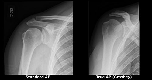
Caption: Why the view matters. The same normal shoulder, imaged two ways. Left: a standard AP, taken straight on relative to the body, where the ball and socket overlap and the joint space is partly hidden. Right: a true AP, or Grashey view, angled to match the plane of the shoulder blade so the beam passes cleanly through the joint. The cartilage space can now be seen directly - which is what allows the extent of arthritis to be assessed rather than estimated.
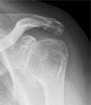
Caption: X-ray of a shoulder with glenohumeral osteoarthritis. The joint space between ball and socket has narrowed, and a bone spur has formed along the lower edge of the humeral head.
CT scan is the key study for surgical planning. It defines the three-dimensional shape of the glenoid, quantifies bone loss, and measures version - how the socket is angled. Glenoid version and bone loss are the two variables that most influence how a shoulder replacement should be performed, and they cannot be assessed accurately on plain X-ray. In my practice, the CT scan also generates the three-dimensional preoperative plan used for computer-navigated arthroplasty. (See: Computer-Navigated Shoulder Replacement)
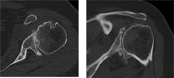
Caption: CT of the same shoulder, looking down through the joint. These slices show the pattern of glenoid wear and how far the socket has tilted backward - detail the X-ray above cannot provide.
Placing these two studies side by side makes the point better than any explanation. The X-ray establishes that arthritis is present. The CT shows the shape of the problem - which way the socket has worn, how far it has rotated, and how much solid bone remains to support an implant. Those are three-dimensional questions, and an X-ray is a two-dimensional image.
MRI is not routinely required once the diagnosis of osteoarthritis is established. X-rays and examination make the diagnosis; an MRI adds little to it and is not something every arthritic shoulder needs.
Where I do find an MRI valuable is when I’m considering an anatomic total shoulder replacement. That implant depends on a rotator cuff capable of supporting it, and I want some assurance the cuff is up to the job before committing to it.
Being precise about what the MRI can and cannot tell us matters here, because its value is asymmetric. An MRI cannot definitively guarantee that a rotator cuff will function well long-term - tendon appearance on imaging and tendon performance under load are related but not the same thing. What it does reliably do is rule patients out. If the MRI shows significant rotator cuff tearing, that patient is not a candidate for an anatomic replacement, and we know it before surgery rather than discovering it in the operating room or, worse, watching the implant fail years later. Identifying who shouldn’t have an anatomic replacement is the practical reason to obtain the study.
Non-Surgical Treatment
Most patients with shoulder arthritis should begin with non-surgical treatment, and many do well with it for years. Surgery is not a race, and no one should feel rushed toward a replacement.
Activity modification. This is not the same as inactivity. It means identifying the two or three specific movements that reliably provoke pain - often overhead reaching or heavy pressing - and finding alternatives. Most patients can stay quite active.
Physical therapy. The goal is preserving motion and maintaining the strength of the muscles around the joint, particularly the rotator cuff and the scapular stabilizers. Therapy will not regrow cartilage, but a well-conditioned shoulder tolerates arthritis considerably better than a deconditioned one. Therapy that aggressively pushes into painful ranges tends to backfire; the program should be tolerable.
Anti-inflammatory medication. Over-the-counter NSAIDs can meaningfully reduce pain. They carry real risks with prolonged use - gastrointestinal, kidney, and cardiovascular - so they should be discussed with your physician rather than taken indefinitely by default.
Corticosteroid injections. A well-placed injection into the glenohumeral joint can provide substantial relief, typically lasting weeks to several months. Because the shoulder joint is deep and the injection needs to reach inside it, image guidance improves accuracy. Injections are useful and also diagnostically informative: if an injection into the joint relieves your pain, that confirms the joint is the pain source. There are limits to how frequently they should be repeated, and an injection shortly before a planned replacement may increase infection risk - timing matters.
Other injectables. Hyaluronic acid injections are used off-label in the shoulder and the evidence is mixed. Platelet-rich plasma and other orthobiologics are an area of genuine ongoing research, but for established, bone-on-bone arthritis, current evidence does not support them as a substitute for definitive treatment. Patients are frequently marketed these treatments at significant out-of-pocket cost, and deserve a candid conversation about what the data actually show.
When Is Surgery the Right Choice?
Shoulder replacement becomes reasonable when arthritis pain interferes with sleep and daily function despite an honest trial of non-surgical care. There is no X-ray finding that mandates surgery. The decision rests on how much the shoulder is limiting your life.
Signals that it is time to have the conversation:
- Pain that consistently wakes you at night
- Progressive loss of motion affecting dressing, grooming, or reaching
- Injections that no longer help, or help for only a short time
- Giving up activities that matter to you
- Bone-on-bone changes on X-ray with symptoms that match
How I Approach This Conversation
When we meet during your visit, I will ask you how much your shoulder is affecting your quality of life. I can’t make the decision for you to have a shoulder replacement, but I do like to give you some guidance on the severity of your symptoms.
Sometimes I will tell you that your function is still good and your pain is minimal, and that I don’t think you would see a meaningful change from a shoulder replacement. Other times I will advise that a shoulder replacement is a reasonable option - because it may offer a more reliable outcome than other treatments that have already failed or no longer work for you.
Both of those are real answers, and which one you hear depends on where you actually are, not on where the X-ray says you are.
Which operation depends primarily on your rotator cuff.
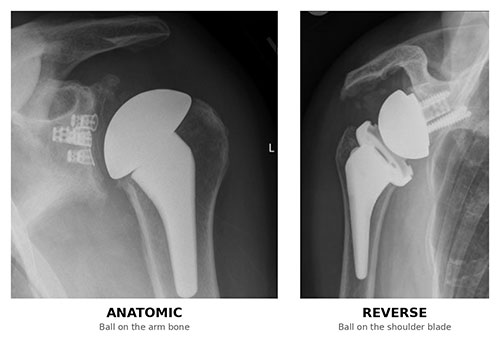
Caption: Anatomic (left) and reverse (right) total shoulder replacement, side by side. In the anatomic replacement the ball sits on the arm bone and the socket on the shoulder blade, as in a normal shoulder. In the reverse, those positions are switched - the ball is fixed to the shoulder blade and the socket to the top of the arm bone.
Anatomic total shoulder replacement resurfaces the ball and socket in their normal configuration. It requires a functioning rotator cuff and, in the right patient, produces excellent pain relief and motion.
Reverse total shoulder replacement switches the positions of ball and socket so the deltoid muscle powers the arm instead of the rotator cuff. It is the appropriate choice when the rotator cuff is deficient, when there is significant glenoid bone loss, and in several other specific situations.
Shoulder arthroscopy has a limited role in true arthritis. It may help selected younger patients with early changes or a mechanically loose body, but it does not treat established arthritis, and patients should be wary of it being offered as a way to delay a replacement that is genuinely indicated.
Why Glenoid Precision Matters
The single most technically demanding part of a shoulder replacement is positioning the socket component. The glenoid is small, often eroded asymmetrically - the B-type pattern described above is the usual reason - and surrounded by limited bone. Malpositioning it is a well-documented cause of early loosening and revision surgery.
This is the reason I perform shoulder arthroplasty using computer navigation on the AdvitaGPS platform. A preoperative CT scan generates a three-dimensional plan, and during surgery the navigation system provides real-time confirmation that the implant is going in exactly where it was planned. In a published analysis of more than 16,000 navigated cases, 98% were completed as planned, with placement verified within approximately 2 mm and 2 degrees of the preoperative plan.
By number of navigated cases performed, I am among the top 5% of surgeons worldwide. (See: Computer-Navigated Shoulder Replacement)
Frequently Asked Questions
Can shoulder arthritis be reversed?
No. Cartilage does not regenerate once it is lost, and no medication, injection, supplement, or exercise program has been shown to reverse established shoulder arthritis. Treatment focuses on controlling pain, preserving motion, and maintaining function - and these goals are very achievable.
How fast does shoulder arthritis progress?
It varies widely. Some patients remain stable for a decade with minimal change; others progress noticeably over two to three years. Post-traumatic arthritis and inflammatory arthritis tend to progress faster than primary osteoarthritis. There is no reliable way to predict an individual’s course, which is why treatment is guided by symptoms rather than by imaging.
Is exercise bad for an arthritic shoulder?
No - the right exercise helps. Maintaining strength and motion improves how well the shoulder tolerates arthritis. What should be modified is heavy overhead loading and movements that reliably produce sharp pain. Swimming, cycling, walking, and appropriately loaded resistance training are generally well tolerated.
Do I need an MRI to diagnose shoulder arthritis?
No - an MRI is not routinely required once osteoarthritis has been diagnosed. Plain X-rays and a physical examination are sufficient to make the diagnosis. An MRI becomes useful when an anatomic total shoulder replacement is being considered, because it helps assess whether the rotator cuff can support that type of implant. A CT scan is obtained when surgery is planned, because it defines glenoid bone shape and version.
Why would my surgeon order an MRI if the X-rays already show arthritis?
Usually to evaluate the rotator cuff before deciding which type of replacement to perform. An anatomic total shoulder replacement relies on a functioning rotator cuff; a reverse replacement does not. An MRI cannot guarantee a rotator cuff will perform well over time, but it can reliably identify significant tearing - which rules out an anatomic replacement. Knowing that before surgery, rather than during it, is the point.
What X-ray views are needed to diagnose shoulder arthritis?
Two views matter most: a true AP (Grashey) view and an axillary view. A standard AP X-ray is taken straight on relative to the body, so the ball and socket overlap and the joint space is partly obscured. A true AP is angled to match the plane of the shoulder blade, letting the beam pass cleanly through the joint so the cartilage space can be seen directly. The axillary view, taken from below, shows whether the humeral head has shifted backward in the socket. Emergency rooms and primary care offices often obtain only a single standard AP, which is appropriate for screening for fracture but may not show the true extent of arthritis.
My X-rays already showed arthritis. Will I need them repeated?
Possibly, and it isn’t duplication for its own sake. If the original films didn’t include a true AP and an axillary view, they may show that arthritis is present without showing how advanced it is or how the socket has worn - and those details determine what treatment is appropriate. Bring any outside imaging to your appointment; it’s always useful, even when additional views are needed.
Should I wait as long as possible before having a shoulder replacement?
Not necessarily. Shoulder replacements are durable, with survivorship commonly reported around 90% at ten years. Waiting until the shoulder is severely stiff, the rotator cuff has failed, or substantial glenoid bone has eroded can make the operation more complex and the outcome less predictable. The reasonable approach is to proceed when symptoms justify it, not to endure years of avoidable disability.
How do I know if I’m ready for a shoulder replacement?
The decision is based on how much the shoulder is affecting your quality of life, not on how bad the X-ray looks. In my practice, that’s the question I’ll ask you directly. If your function is still good and your pain is minimal, I’ll usually say I don’t think a replacement would give you a meaningful change. If other treatments have failed or stopped working, I’ll tell you a replacement is a reasonable option because it tends to offer a more reliable outcome. The decision itself is yours - my job is to give you an honest read on where you stand.
Can shoulder arthritis cause pain down my arm?
It can cause pain referred to the outer upper arm, but pain that travels past the elbow into the forearm or hand - particularly with numbness, tingling, or weakness - suggests a nerve source in the neck rather than the shoulder joint. This distinction is important and is covered in detail here.
Ready to talk about your shoulder?
Dr. Gabriella Ode is a fellowship-trained shoulder and sports medicine surgeon at the HSS Sports Medicine Institute, with offices on the Upper East Side, the West Side, and in Brooklyn. Request an appointment or request a remote second opinion if you’re outside the New York area.
Financial disclosure.
Dr. Ode is a paid consultant for Advita Ortho and ConMed, manufacturers of orthopaedic implants and surgical instrumentation. These relationships are reported publicly through the CMS Open Payments database. She receives no compensation for this website, and no compensation is tied to any individual patient’s treatment decision. Her complete financial disclosures are available here.
This article is for general education and is not a substitute for individual medical advice. Please consult a physician about your specific condition.