Frozen Shoulder (Adhesive Capsulitis): Stages, Timeline, and What Actually Helps
Frozen shoulder is a condition in which the capsule surrounding the shoulder joint becomes inflamed, thickened, and contracted, causing severe pain followed by profound stiffness. Its defining feature is that motion is lost in every direction - not only when you move the arm yourself, but also when someone else moves it for you. That single characteristic is what separates it from almost every other shoulder problem.
Lifetime prevalence is estimated at 2–5% of the general population. The peak age of onset is around the mid-50s, but the condition is common from the forties onward, and it is more frequent in women - roughly 1.4 women for every man. The non-dominant shoulder is affected more often than the dominant one.
In my practice, a substantial proportion of these patients are women in their forties, and the overlap with perimenopause is difficult to ignore. That observation has support in the research: estrogen influences collagen structure and turnover, and declining estrogen is associated with the kind of connective-tissue changes seen in this condition. A 2023 study from Duke found that women not taking menopausal hormone therapy were roughly twice as likely to develop frozen shoulder. Whether hormone therapy has a role in treating frozen shoulder is a separate question and one that remains under investigation - a trial at UCSF is examining it as an adjunct treatment in peri- and postmenopausal women.
Why this matters practically, rather than as an interesting aside. If you are a woman in your forties or early fifties with a shoulder that has become painful and stiff without an injury, frozen shoulder belongs high on the list - and it is frequently not where the first evaluation lands. It also means that if you are already discussing perimenopausal symptoms with a gynecologist or primary care physician, mention your shoulder in the same conversation. The two are more connected than either specialty has historically treated them.
It is among the most difficult conditions in orthopaedics to live through, largely because the timeline is long and the early phase is severely painful. It is also, for most patients, a condition that improves substantially - around 80% regain near-normal or normal shoulder function with appropriate treatment.
What’s Actually Happening Inside the Shoulder
The shoulder joint is enclosed in a capsule - a sleeve of connective tissue that is normally loose and elastic, with generous folds that allow the considerable range of motion the shoulder is capable of.
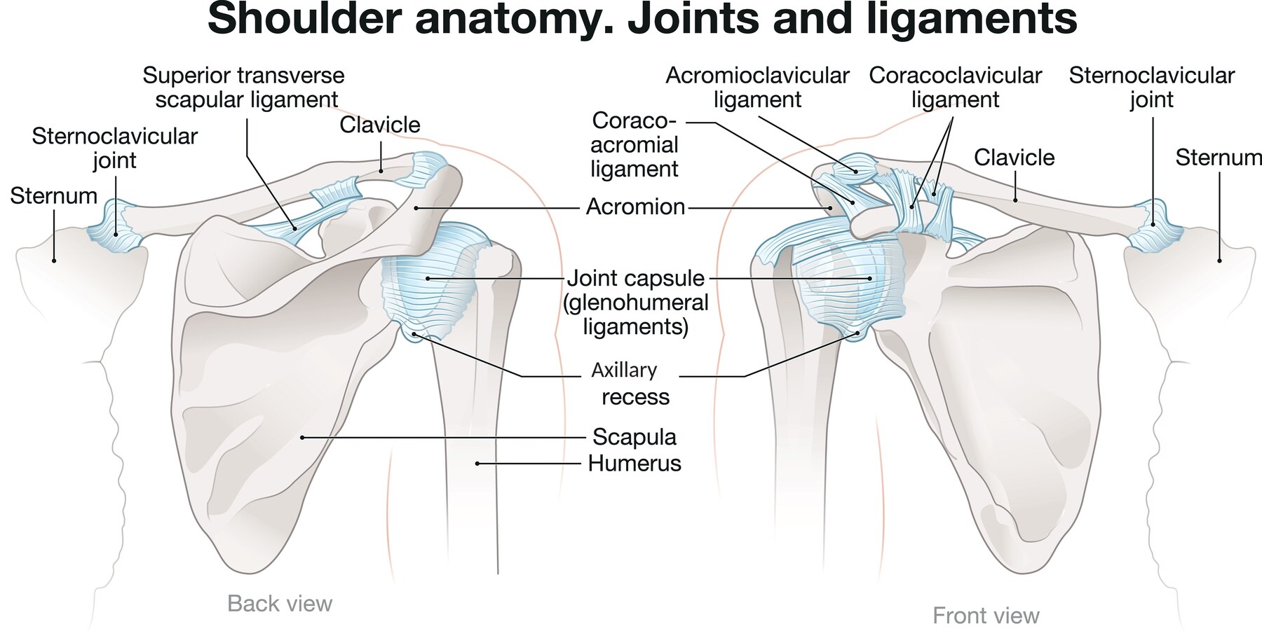
Caption: The normal shoulder joint capsule, seen from behind (left) and from the front (right). The capsule is the pale blue sleeve enclosing the joint. Note the axillary recess at its lowest point — a deliberate fold of slack tissue that unfurls as you raise your arm. A healthy capsule has redundancy built into it, which is what allows the shoulder its range of motion. Adhesive capsulitis is, in essence, the loss of that redundancy.
Look at the axillary recess on that illustration, because it explains a great deal about this condition. It is a pouch of deliberately loose capsule at the bottom of the joint. When the arm hangs at the side it is folded and redundant; when the arm elevates, it unfolds. In adhesive capsulitis that recess contracts and obliterates, and with it goes the slack the shoulder relies on.
In adhesive capsulitis, this capsule becomes inflamed and then fibrotic. Inflammatory cells infiltrate the tissue, new blood vessels and nerve fibers grow into it (which is why the early phase is so painful), and the capsule progressively thickens and contracts. The rotator interval and the coracohumeral ligament at the front of the shoulder are typically affected first and most severely, which is why loss of external rotation is usually the earliest and most pronounced restriction.
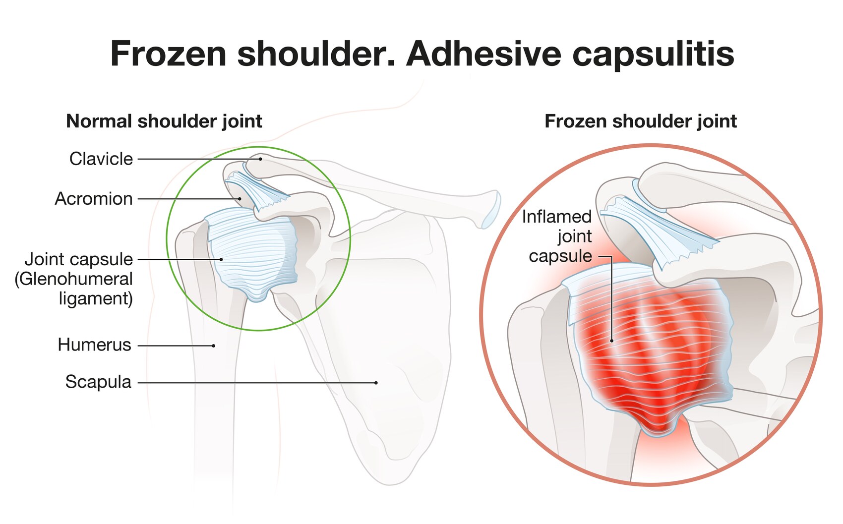
Caption: A normal joint capsule (left) compared with the inflamed, thickened capsule of adhesive capsulitis (right). In the affected shoulder the capsule is not merely tight — it is inflamed, thickened, and contracted, with the normal folds of slack tissue lost. This is why the condition is so painful in its early phase and so stiff in its later one, and why the two problems arrive in that order.
The capsule physically shrinks. This is why stretching alone is slow to help, and why forcing motion early makes matters worse. You are not stretching a tight muscle - you are pulling against inflamed, contracting scar tissue, and that tissue responds to being forced by becoming more inflamed.
Who Gets It
Primary (idiopathic) frozen shoulder arises without an identifiable cause. It is the most common form.
Secondary frozen shoulder follows a triggering event - a period of immobilization after injury or surgery, a minor injury that caused the arm to be protected, or another shoulder condition such as a rotator cuff tear, a proximal humerus fracture, or arthritis. It also occurs after shoulder surgery, including rotator cuff repair.
This distinction is not academic - it changes the treatment timeline. A primary frozen shoulder is generally given a long runway, because it improves on its own in most people. A secondary one, where stiffness is obstructing recovery from something else, may warrant earlier intervention. The treatment section below sets out how I approach each.
Established risk factors:
- Diabetes - the strongest association by a wide margin. Reported incidence in people with diabetes reaches close to 60% in some series, and the course tends to be more severe, more prolonged, and more likely to involve both shoulders. Longer duration of diabetes is associated with poorer treatment outcomes. If you have frozen shoulder and have not been screened for diabetes, you should be.
- Thyroid disease, both hyperthyroidism and hypothyroidism
- Female sex, particularly from the forties onward - see the note on perimenopause above
- Immobilization after any injury or surgery
- Cardiovascular disease, Parkinson’s disease, stroke, and Dupuytren’s disease - the last of these is a fibrotic condition of the palm with a strikingly similar underlying biology
- Raised cholesterol, smoking, obesity, and low physical activity levels
Frozen shoulder is rarely an isolated finding. More than 80% of people diagnosed with it have at least one of these associated conditions, and over a third have three or more. That pattern is part of why it is better understood as a systemic condition that manifests in the shoulder than as a purely local problem.
The other shoulder. Around 17% of patients develop frozen shoulder on the opposite side within five years. Recurrence in the same shoulder, by contrast, is uncommon.
The Three Stages
Frozen shoulder progresses through three stages, and the untreated natural course runs somewhere between one and three years. But that figure describes the condition left to run its course - it is not a prediction for every patient, and how early it is caught makes a substantial difference.
Caught early, this can be a much shorter story. A frozen shoulder identified in the painful inflammatory phase, before significant stiffness has set in, often responds well to an intra-articular injection combined with physical therapy - and can settle within a few months rather than a few years. A shoulder that is already profoundly stiff by the time it is diagnosed takes considerably longer.
Several things influence which of those courses you are on:
- How quickly the diagnosis is made. This is the single biggest lever, and it is why recognizing the freezing stage below matters so much.
- Systemic factors, diabetes in particular. These tend to lengthen the course regardless of what treatment is given.
- Access to consistent, good-quality physical therapy. Not simply a referral - sustained, competent therapy.
- Your consistency with the home exercise program. Frequent short sessions do more than occasional intensive ones, and this is the part of the process you control.
The stages below describe the shape of the condition, not a fixed schedule.
Stage 1 - Freezing (painful stage). Roughly 2 to 9 months. Pain dominates. It builds gradually, becomes severe, and is characteristically worst at night - many patients cannot sleep on that side and are woken repeatedly. Motion begins to decline, but pain is the main complaint.
This is the stage where the diagnosis is most often missed, because early on it resembles a rotator cuff problem. It is also the stage where treatment does the most good. A shoulder caught here, before the capsule has fully contracted, has the best chance of a short course. If you have unexplained shoulder pain that is worse at night and your arm is starting to feel restricted, have that combination examined rather than waiting to see whether it settles.
Stage 2 - Frozen (stiff stage). Roughly 4 to 12 months. Pain gradually improves, sometimes substantially. Stiffness is now the dominant problem. The shoulder is markedly restricted in all directions, and daily tasks - reaching a seatbelt, fastening a bra, washing hair, putting on a coat - become genuinely difficult. Many patients say this stage is more functionally limiting but far more tolerable than the first.
Stage 3 - Thawing (recovery stage). Can last 12 months or longer. Motion returns gradually. Recovery is usually slow and steady rather than dramatic. Most patients regain most or all of their motion.
Two qualifications, stated plainly.
First, the staging is a useful framework rather than a precise timetable. The stages overlap, the durations vary considerably between individuals, and the classification itself is debated - many patients continue to have symptoms and functional restriction well beyond the period the model predicts. It is best used to describe which problem is dominant rather than to predict a date.
Second, the traditional teaching that frozen shoulder is entirely self-limiting is more optimistic than the evidence supports. Reported rates of persistent pain or stiffness in the long term vary widely depending on the study and on how “persistent” is defined - from around 10–20% in some series to as high as 20–50% in others. For most people the residual symptoms are mild and functionally unimportant. For a minority they are not. You should have that range, because a blanket assurance that the condition always resolves completely sets an expectation that is not always met.
Frozen Shoulder or Rotator Cuff Tear?
These two conditions are mistaken for one another frequently, particularly early on, and the distinction becomes straightforward once you know what to test.
The decisive test is passive motion.
| Frozen shoulder | Rotator cuff tear | |
|---|---|---|
| Passive motion (someone else moves your arm) | Severely limited - this is the key finding | Usually preserved |
| Active motion | Severely limited | Limited by pain or weakness |
| External rotation with elbow at side | Markedly restricted - the hallmark | Usually normal |
| Strength when tested in available range | Normal | Weak |
| Onset | Gradual, often no injury | May follow an injury |
| Pain pattern | Severe, diffuse, worst at night | Outer upper arm, worse overhead |
Loss of passive external rotation with the elbow at the side is the single most useful sign. A rotator cuff tear does not restrict it. Frozen shoulder almost always does.
Motion is typically lost in a characteristic order, which is itself a useful diagnostic clue: external rotation first, then abduction, then internal rotation, and flexion last. A shoulder that has lost external rotation before anything else, without an injury to explain it, is behaving like a frozen shoulder.
Why this distinction needs settling before any treatment begins. Rotator cuff abnormalities are extremely common on imaging - present in 96% of shoulders with no symptoms at all in a general-population study - so an MRI in a patient over 50 will frequently show a tear whether or not it has anything to do with why the shoulder hurts. If the shoulder is also frozen, both findings are real, and the examination is what establishes which one is producing the symptoms.
In practice this means the stiffness is usually addressed first. A capsule that has contracted will keep the shoulder restricted regardless of what the tendons look like, and a frozen shoulder that settles often takes the pain with it. Where a tear does turn out to be contributing, it can be reassessed once motion has returned - by which point the picture is far clearer than it was at the start.
If the picture remains unclear, the differential is wider still. See the page entitled “Is Your Shoulder Pain Actually Coming From Your Neck?”, and the page on scapular dyskinesis.
Diagnosis
Frozen shoulder is a clinical diagnosis made by physical examination. No imaging test is required to make it.
X-rays are typically obtained to exclude arthritis, which can cause similar stiffness - and in frozen shoulder, X-rays are normal. Blood work to screen for diabetes and thyroid disease is reasonable given how strong those associations are.
Where an MRI fits. An MRI is not required to make the diagnosis, and a scan is never the reason I conclude that a shoulder is frozen - the examination establishes that. But there are several situations in which I do order one, and the reasons matter, because “you don’t need an MRI to diagnose this” and “I would like an MRI” are not in conflict:
- To support the diagnosis. Adhesive capsulitis has recognizable features on MRI - thickening of the capsule, changes at the rotator interval and the coracohumeral ligament, and loss of the normal axillary recess. Where the clinical picture is atypical, seeing those findings is reassuring.
- To establish a baseline before other treatment. This is the reason I most often order one. Frozen shoulder frequently occurs in a shoulder that has other wear and tear in it, and it is useful to know what else is present before starting down a treatment path - particularly before an injection, a hydrodilatation, or any consideration of surgery. It changes what I plan for and what I tell you to expect.
- To exclude another problem where the presentation does not fit cleanly.
The distinction that matters is what the scan is being used for. It is not being used to establish that your shoulder is frozen. It is being used to understand the whole shoulder before treating it - and to make sure that when motion returns, we already know whether there is anything else that will need attention.
A point about where an injection is placed, because it matters more here than patients realize. The shoulder has two separate spaces that can be injected: the subacromial space, above the rotator cuff, and the glenohumeral joint itself, inside the capsule. Injections for rotator cuff problems and bursitis are placed in the subacromial space. For adhesive capsulitis I place the injection into the joint, because an injection inside the capsule concentrates the medication on the capsule itself - which is the tissue that is inflamed and contracted in this condition.
This matters if you have already had a shoulder injection that did not help. It does not mean injections will not work for you. It may simply mean the medication was delivered to a different compartment than the one producing your symptoms.
A related point on numbing. If local anesthetic settles the pain but the shoulder still will not move, that is informative - it means the restriction is mechanical rather than driven by pain, which is characteristic of a truly frozen shoulder.
How I Work Out Which Stage You Are In
Staging matters more here than in almost any other shoulder condition, because it determines what I do next. The distinction I am making is not really about how many weeks it has been - patients rarely date the onset accurately, and the stages overlap. It is about which problem is dominant.
If pain is the dominant complaint - pain at rest, pain at night, pain that arrives before the end of the available range rather than at the end of it - you are in the inflammatory phase, whatever the calendar says. The priority is settling that down. Pushing range in this phase tends to prolong it.
If stiffness is the dominant complaint - the shoulder is restricted, the restriction is what limits you, and the pain is mostly the pain of hitting the end of the range - the inflammatory phase is passing and motion work becomes the priority. This is when therapy earns its keep and when it can be progressed more assertively.
The most useful question I ask is where in the range the pain occurs. Pain throughout the range, and at rest, is inflammation. Pain only at the end of the range is a mechanical block. Those two findings call for different treatment, and the answer is usually clear within a minute of examining the shoulder.
Treatment
Treatment is stage-dependent, and matching the treatment to the stage is what determines whether it helps. In the painful stage, the priority is controlling inflammation rather than stretching. In the stiff stage, the priority shifts to restoring motion. The same intervention applied at the wrong point in the course can be unhelpful or actively counterproductive - which is why the first question is always where in the process you are.
Corticosteroid injection - the highest-value early intervention. An intra-articular corticosteroid injection during the inflammatory stage has the strongest evidence of any treatment for improving the early course. It is more effective than placebo, and more effective than physical therapy alone for pain in the early stages. The benefit is greatest when given early, before capsular contraction is established - injections given in stage I or II produce considerably more improvement than those given later.
Two honest qualifications. The advantage over physical therapy narrows considerably by six and twelve months, so an injection accelerates the early course rather than changing the eventual destination. And an injection on its own does little for range of motion - it is the combination of injection and therapy that improves movement. That is precisely how it should be used: the injection creates the window, and the therapy uses it.
If you have diabetes, be aware that an intra-articular corticosteroid injection can cause a transient rise in blood glucose for several days afterwards. It is manageable and it is not a reason to avoid the injection, but anticipate it and mention it to whoever manages your diabetes.
Hydrodilatation (capsular distension). Also written hydrodilation - the two spellings are used interchangeably for the same procedure, so you may see either.
An image-guided injection of a larger volume of sterile fluid, usually with corticosteroid, that distends and mechanically stretches the contracted capsule. Meta-analysis supports it: hydrodilatation with corticosteroid produces superior short-term pain relief, range of motion, and function compared with placebo, with motion gains persisting beyond 24 months - and it has been found to offer greater benefit than corticosteroid injection alone. Among the alternatives to standard injection, it has some of the strongest supporting evidence.
How I use it. Hydrodilatation is a common adjunct in my practice, and I refer patients out for it rather than performing it myself. It is done under ultrasound or fluoroscopic guidance. In my practice the colleagues who perform it are most often primary care sports medicine physicians, though depending on the setting it may also be done by a physiatrist or a musculoskeletal radiologist.
When I refer. My usual threshold is failure of one or more intra-articular corticosteroid injections. If the injection has not produced enough improvement, or the improvement has not lasted, hydrodilatation is the next step I reach for before considering anything surgical.
What it involves varies. The procedure may be performed on its own, or combined with a nerve block, depending on the preference of the physician performing it and on what you have already had done. That is a reasonable variation in practice rather than a difference in quality, and the physician doing the procedure is the best person to explain which approach they are using and why.
A word on who performs these procedures, because the range of specialties can be confusing. Non-operative musculoskeletal care is provided by several different kinds of physician - primary care sports medicine, physical medicine and rehabilitation (physiatry), and musculoskeletal radiology among them. The useful question is not the specialty label but whether the physician performs the procedure regularly and under image guidance. For a hydrodilatation, accurate placement inside the joint is the entire point, and that comes from experience with the procedure rather than from any one training pathway.
One point of reassurance if you are referred to “sports medicine” for this: a primary care sports medicine physician is a non-surgical specialist. You are not being sent to another surgeon.
Physical therapy - with the right intensity. Gentle, pain-limited range of motion work is helpful throughout. Aggressive stretching during the painful stage is counterproductive and can prolong the inflammatory phase. Therapy becomes more valuable, and can be pushed harder, once pain has settled and the shoulder is in the stiff stage. A home program done consistently in short frequent sessions outperforms occasional aggressive sessions.
Oral medication. NSAIDs help with pain.
A short course of oral corticosteroid is a reasonable option in early frozen shoulder, and it is one I offer to patients who would rather not begin with an injection into the joint. Not everyone is comfortable starting with a needle in the shoulder, and that preference does not have to mean going without treatment during the phase when treatment does the most good.
What to expect from it, honestly. Oral steroid produces meaningful short-term improvement in pain for many patients. Its limitations are that the benefit is not sustained as reliably as an intra-articular injection, and some patients experience rebound pain when the tablets finish. Where it works well, it can do the same essential job - settling the inflammation enough to allow productive physical therapy - without an injection.
It is not an indefinite treatment, and it is less suitable for some patients, including those with diabetes, in whom oral steroid raises blood glucose more substantially and for longer than a single joint injection does. If a short oral course does not achieve enough, an intra-articular injection remains available as the next step.
Surgery - for the minority who don’t improve. My threshold differs depending on whether the frozen shoulder is primary or secondary, and this distinction matters more than the calendar alone.
For primary (idiopathic) frozen shoulder - the kind that arises on its own - I wait more than twelve months before considering surgical intervention. This is a condition that improves with time in most people, the natural course is long, and operating early means operating on shoulders that would have improved anyway. Patience here is a clinical position, not a delay.
For secondary frozen shoulder, I will sometimes intervene earlier. The situations where I consider it are ones in which the stiffness is obstructing something else that needs to progress:
- Stiffness following a rotator cuff tear
- Adhesive capsulitis that develops after rotator cuff repair and is slowing the rehabilitation
In each of these the shoulder is not simply waiting out a self-limiting condition - the stiffness is actively interfering with recovery from something else, and the cost of waiting is higher. Even then, injections come first, and I move toward surgery only when they have not resolved the problem.
Two techniques exist, and they are often discussed as alternatives:
- Manipulation under anesthesia (MUA) - the capsule is stretched and released while you are asleep. Reported outcomes are good in more than 80% of patients.
- Arthroscopic capsular release (ACR) - the contracted capsule is divided under direct vision through small incisions, typically at the rotator interval and along the front and inferior capsule.
What I Do, and Why
I perform both at the same operation - a manipulation combined with an arthroscopic capsular release. They are not competing options in my hands; each does something the other does not.
The arthroscopy lets me see the joint. Before releasing anything, I can perform a diagnostic arthroscopy and look at what is actually there - the state of the capsule, and whether anything else in the joint is contributing. That matters in a condition where imaging is not definitive and where a stiff shoulder can have more than one thing going on inside it.
It also allows the release itself to be precise. Rather than relying on stretching alone to tear the capsule wherever it happens to give way, the adhesed capsule is divided deliberately, where it is actually contracted, under direct vision. A manipulation applies force and the tissue fails where it fails; a release under vision decides where. Combining the two means the manipulation is completing a release that has already been directed rather than doing the work blind.
There is a safety argument as well. The risks associated with forceful manipulation - humeral fracture in particular, and less commonly rupture of the biceps or subscapularis tendon - arise from the force required when the capsule has not been released first. Releasing under vision reduces the force needed. Those risks matter most in patients with osteoporosis, which is a relevant consideration in exactly the age group this condition affects.
What the largest trial found. UK FROST is the biggest randomized comparison in this condition, and it compared early structured physiotherapy, MUA alone, and arthroscopic release. At twelve months, most patients in all three groups had improved to near-normal function. Arthroscopic release produced the best scores and required the least additional treatment afterwards; manipulation outperformed physiotherapy alone. But the differences were modest, and the trial’s conclusion was that all three approaches produced substantial recovery.
The reasonable reading is that surgery is not a shortcut to be offered early - it belongs after less invasive measures, which is why the timeline above matters. Once that point is reached, the arthroscopic component is the one associated with the most durable result.
Either way, immediate post-operative therapy is essential. The motion gained in the operating room is easily lost in the first two weeks if therapy does not begin straight away. This is the part of the operation the patient controls, and it determines much of the outcome.
Patients with diabetes tend to respond more slowly and less completely to every treatment on this list. You should know that at the outset rather than encounter it as an unexplained disappointment three months in - it does not mean treatment will not work, but the timeline is often longer and expectations should be set accordingly.
Frequently Asked Questions
How long does frozen shoulder last?
It depends substantially on how early it is diagnosed. Left to run its natural course, frozen shoulder takes somewhere between one and three years - the freezing stage roughly 2 to 9 months, the frozen stage roughly 4 to 12 months, and the thawing stage 12 months or more. But a frozen shoulder caught early, in the painful phase before significant stiffness has set in, often responds well to an intra-articular injection combined with physical therapy and can settle within a few months. A shoulder that is already profoundly stiff at diagnosis takes considerably longer.
Other factors that lengthen recovery include diabetes and other systemic conditions, access to consistent good-quality physical therapy, and how consistently the home exercise program is done. The published stage durations describe the shape of the condition rather than a schedule your shoulder is obliged to follow.
Does frozen shoulder always go away on its own?
Most cases improve substantially - around 80% of patients regain near-normal or normal function with appropriate treatment. But not every case resolves completely. Reported rates of lasting pain or stiffness range from about 10–20% in some studies to as high as 20–50% in others, depending on how persistence is defined. For most people any residual symptoms are mild. Treatment during the painful stage, particularly a corticosteroid injection combined with therapy, improves comfort and shortens the difficult phase.
What’s the fastest way to treat frozen shoulder?
The strongest evidence supports an intra-articular corticosteroid injection early in the inflammatory stage, combined with gentle, pain-limited range of motion work. The injection alone does little for motion; it is the combination that works. For patients who remain stiff, hydrodilatation with corticosteroid improves pain, motion, and function, with gains that can persist beyond two years. Aggressive stretching during the painful phase tends to prolong symptoms rather than shorten them.
Should I push through the pain when stretching a frozen shoulder?
No - not during the painful freezing stage. Forcing motion against an actively inflamed capsule can worsen inflammation and extend that phase. Stretching should be gentle and stay within a tolerable range. Once pain has settled and stiffness is the main issue, therapy can be progressed more assertively.
Why do people with diabetes get frozen shoulder more often?
The association is well established and thought to relate to glycation of collagen, which alters the structure of connective tissue and makes the capsule more prone to fibrosis. People with diabetes have a substantially higher lifetime incidence, often more severe and prolonged courses, and a higher likelihood of both shoulders being affected.
Can frozen shoulder come back?
Recurrence in the same shoulder is uncommon. The opposite shoulder is a different matter - around 17% of patients develop frozen shoulder on the other side within five years.
Is frozen shoulder related to menopause?
There appears to be a real connection. Frozen shoulder is most common in women between about 40 and 60, which overlaps closely with the perimenopausal transition. Estrogen influences collagen structure and turnover, and declining estrogen is associated with changes in connective tissue of the kind seen in this condition. A 2023 Duke study found that women not taking menopausal hormone therapy were roughly twice as likely to develop frozen shoulder. Whether hormone therapy can treat frozen shoulder is a different question, and one that remains under investigation - a trial at UCSF is examining it as an adjunct treatment in peri- and postmenopausal women. The association is not the same as an established treatment, so this is not a reason to start hormone therapy for your shoulder. It is a reason, if you are in your forties or fifties with an unexplained stiff and painful shoulder, to raise the connection with both your shoulder specialist and whoever manages your menopausal care.
Where should a cortisone injection be placed for frozen shoulder?
Into the glenohumeral joint itself rather than the subacromial space above the rotator cuff. The two are separate compartments. Adhesive capsulitis is a condition of the joint capsule, so an injection placed inside the joint concentrates the medication on the tissue that is actually affected. Injections into the subacromial space are used for rotator cuff and bursal problems. If you have had a shoulder injection that did not help, find out which compartment it went into.
Is there an option other than an injection for early frozen shoulder?
Yes. A short course of oral corticosteroid is a reasonable alternative in the early inflammatory phase, and Dr. Ode offers it to patients who would rather not begin with an injection into the joint. It produces meaningful short-term pain improvement for many patients, though the benefit is generally less sustained than an intra-articular injection and some patients get rebound pain when the tablets finish. The purpose is the same either way - settling the inflammation enough to allow productive physical therapy. Oral steroid is less suitable for patients with diabetes, in whom it raises blood glucose more substantially and for longer than a single joint injection. If a short oral course does not achieve enough, an injection remains available as the next step.
Does a corticosteroid injection affect blood sugar if I have diabetes?
It can. An intra-articular corticosteroid injection may cause a transient rise in blood glucose for a few days afterwards. It is manageable and it is not a reason to avoid an injection that would help, but anticipate it and mention it to whoever manages your diabetes so you can monitor more closely for a short period.
What is hydrodilatation (or hydrodilation), and when is it used?
Hydrodilatation - also spelled hydrodilation, the two terms are interchangeable - is an image-guided injection of a larger volume of fluid, usually with corticosteroid, that distends and stretches the contracted joint capsule. Evidence supports it: it improves pain, motion, and function compared with placebo, with motion gains that can persist beyond two years, and it has been found to offer greater benefit than a standard corticosteroid injection alone. Dr. Ode refers patients for hydrodilatation as a common next step after one or more intra-articular corticosteroid injections have not produced lasting improvement. It is performed under image guidance - in her practice most often by a primary care sports medicine physician, and in other settings by a physiatrist or a musculoskeletal radiologist. It may be done on its own or combined with a nerve block, depending on the physician performing it and on what has already been tried.
How long before surgery is considered for frozen shoulder?
It depends on whether the frozen shoulder is primary or secondary. For primary (idiopathic) frozen shoulder - the kind that arises without a clear cause - Dr. Ode waits more than twelve months before considering surgical intervention, because the condition improves with time in most people and operating early means operating on shoulders that would have recovered anyway. For secondary frozen shoulder, where stiffness follows a rotator cuff tear or develops after rotator cuff repair and is obstructing rehabilitation, earlier intervention is sometimes appropriate. Injections are still tried first in those cases.
Is surgery better than physical therapy for frozen shoulder?
Not straightforwardly. The largest randomized trial in this condition compared early structured physiotherapy, manipulation under anesthesia, and arthroscopic capsular release. At twelve months most patients in all three groups had improved to near-normal function. Arthroscopic release produced the best scores and required the least further treatment, but it is also the most invasive, which is why it is reserved for patients who have not improved with less invasive measures rather than offered first.
Manipulation or arthroscopic release - which is better?
Dr. Ode performs both at the same operation rather than choosing between them. The arthroscopy allows a diagnostic look inside the joint before anything is released, which matters in a condition where imaging is not definitive, and it allows the contracted capsule to be divided precisely under direct vision rather than torn wherever it happens to give way under stretching. Releasing the capsule first also reduces the force required for the manipulation, which lowers the risk of the complications associated with manipulation alone - humeral fracture in particular, and less commonly tendon rupture.
Do I need an MRI to diagnose frozen shoulder?
Not to make the diagnosis. Frozen shoulder is diagnosed on examination, and the finding that establishes it - loss of passive external rotation with the elbow at the side - cannot be seen on a scan. X-rays are usually obtained to exclude arthritis and are normal in frozen shoulder. That said, an MRI is often reasonable for other purposes: to support the diagnosis where the presentation is atypical, to exclude another problem, and - most commonly - to establish a baseline of what else is going on in the shoulder before starting injections or considering a procedure. Adhesive capsulitis does have recognizable MRI features, including capsular thickening and loss of the normal axillary recess. One thing to be aware of: an MRI in a patient over 50 will often also show a rotator cuff tear, which may or may not be contributing to the symptoms.
Can frozen shoulder develop after shoulder surgery?
Yes, and this is one of the recognized forms of secondary frozen shoulder. Any period of immobilization can trigger it, which is part of why post-operative protocols specify when motion should begin rather than leaving it open-ended. If your shoulder is becoming stiffer rather than looser as you progress through a rehabilitation program, raise it promptly rather than waiting for the next scheduled visit.
Is it safe to do physical therapy during the painful stage?
Yes, provided it is the right kind. Gentle, pain-limited range of motion work is appropriate throughout and helps maintain what motion you have. What is counterproductive in the painful stage is aggressive stretching into pain, which tends to inflame the capsule further and prolong that phase. The rule of thumb is that therapy should not leave you in more pain hours afterwards than when you started.
Is frozen shoulder the same as a rotator cuff tear?
No. The defining difference is passive motion. In frozen shoulder, motion is severely limited even when someone else moves your arm, and external rotation with the elbow at your side is markedly restricted. With a rotator cuff tear, passive motion is usually preserved and the main finding is weakness.
Also on this topic: Dr. Ode contributed expert commentary on frozen shoulder to SELF magazine.
Struggling with a stiff, painful shoulder? Dr. Gabriella Ode is a fellowship-trained shoulder and sports medicine surgeon at the HSS Sports Medicine Institute, with offices in Manhattan and Brooklyn. Frozen shoulder is highly treatable, and treatment works best when matched to the stage you’re in. Request an appointment or request a remote second opinion.
References
- Li D, St Angelo JM, Taqi M. Adhesive Capsulitis (Frozen Shoulder). StatPearls. Updated March 2025.
- Kim J-Y, Gahlot N, Park HB. Frozen shoulder: a narrative review of current treatment concepts and the underlying scientific evidence. Clin Shoulder Elbow. 2025;28(4):529–546.
- Frozen shoulder. Nature Reviews Disease Primers.
- Wittstein J, et al. Duke University School of Medicine - association between menopausal hormone therapy and adhesive capsulitis, presented 2023. Duke Department of Orthopaedic Surgery summary
- Hormone Replacement Therapy as an Adjunct Treatment for Adhesive Capsulitis of the Shoulder in Peri- and Postmenopausal Women. UCSF, NCT07278323 - ongoing.
- Rangan A, et al. UK FROST - multicentre randomized comparison of early structured physiotherapy, manipulation under anaesthesia, and arthroscopic capsular release.
Financial disclosure.
Dr. Ode is a paid consultant for Advita Ortho and ConMed, manufacturers of orthopaedic implants and surgical instrumentation. These relationships are reported publicly through the CMS Open Payments database. She receives no compensation for this website, and no compensation is tied to any individual patient’s treatment decision. Her complete financial disclosures are available here.
This article is for general education and is not a substitute for individual medical advice. Please consult a physician about your specific condition.