Anterior Shoulder Instability: Why It Happens and Why It Recurs
Anterior shoulder instability means the ball of the shoulder slips forward out of its socket - completely, which is a dislocation, or partially, which is a subluxation. It accounts for the large majority of shoulder dislocations and is overwhelmingly a young person’s problem, concentrated in the second and third decades of life and in contact and collision athletes.
Two facts shape almost every decision that follows.
After a first traumatic dislocation in a young athlete, the shoulder is very likely to dislocate again without surgery. Recurrence in conservatively managed patients under twenty commonly exceeds 70 percent.
Every dislocation raises the likelihood of the next one. Each episode stretches the capsule and ligaments a little further, so the force required to displace the shoulder tends to fall over time. Some patients reach that point with the bone still intact. Where bone loss does develop - on the socket or the humeral side - the process accelerates, because a rim that has lost bone contains the ball less securely.
For treatment, see: Treating Anterior Shoulder Instability
Dislocation, Subluxation, and Direction
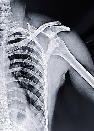
Caption: An anterior shoulder dislocation on X-ray. The ball of the shoulder is no longer sitting in its socket - it has moved forward and downward, coming to rest below and toward the midline. On a normal film the ball sits centered against the socket. This is what the injury looks like before the shoulder is put back in.
- A dislocation is a complete separation. The ball comes fully out and usually has to be put back in
- A subluxation is a partial slip that reduces on its own - described as the shoulder “going out and coming back,” a sudden dead feeling in the arm, or a sensation of shifting. Subluxations can cause the same structural damage as a full dislocation, so a shoulder that has never formally dislocated can still be unstable
Direction determines treatment:
- Anterior - forward, and by far the most common. Almost always traumatic. This page
- Posterior - backward. Frequently presents as pain with pushing rather than any sense of the shoulder coming out
- Multidirectional - looseness in more than one direction, usually without significant injury. Treated almost oppositely, which is why separating the two is the first job of the assessment
What Tears When a Shoulder Dislocates
The socket is shallow - the usual comparison is a golf ball on a tee. Stability comes almost entirely from soft tissue: the labrum, the capsule and ligaments, and the dynamic control of the rotator cuff and scapular muscles.
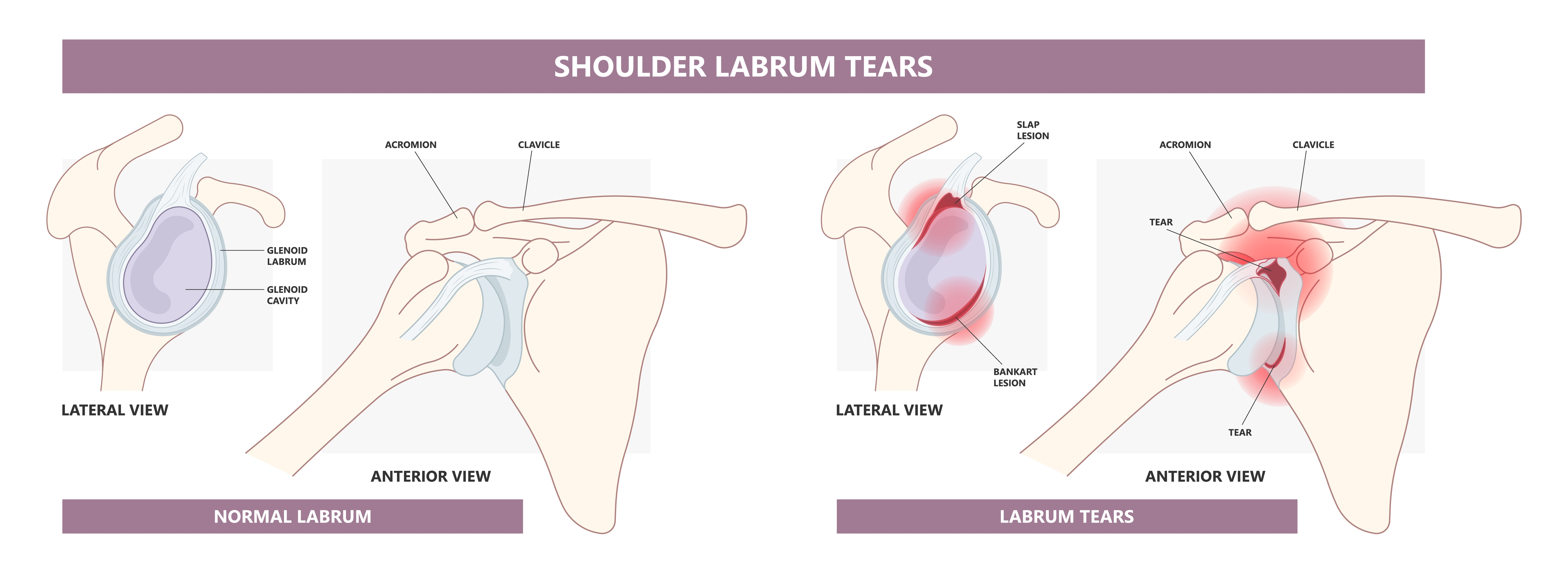
Caption: A normal labrum, and a torn one. Left: the labrum is the rim of cartilage around the edge of the socket, from the side and from the front. Right: the two most common tear locations. A Bankart lesion is at the front rim - the injury that occurs when the shoulder dislocates forward. A SLAP lesion is at the top, where the biceps anchors, and is a different problem.
A predictable pattern of damage follows a forward dislocation:
- Bankart lesion. The labrum and attached ligaments tear off the front rim. This is the essential lesion, present in the great majority of first-time traumatic dislocations. Once detached, the labrum often heals in a stretched or displaced position, leaving the front of the shoulder permanently less secure
- Hill-Sachs lesion. As the humeral head rides over the front rim, the rim stamps a dent into the back of the head. Small ones are of little consequence; large ones can catch on the rim and lever the shoulder out again
- Bony Bankart and glenoid bone loss. The front rim can fracture at the time of injury or erode with repeated episodes. This is the finding that most changes the surgical plan
- Capsular stretching, which adds a further element any repair has to address
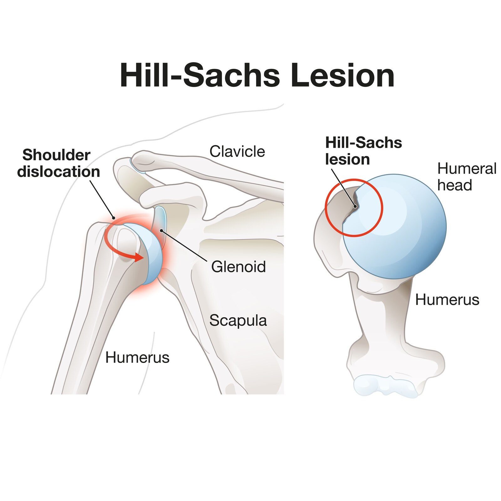
Caption: How a Hill-Sachs lesion forms. Left: as the shoulder dislocates, the ball is driven forward across the front rim of the socket. Right: the rim presses a dent into the back of the ball where the two made contact. Whether that dent matters depends on its size and position - which is what the glenoid track, described below, is used to assess.
Where the Tear Sits: The Clock Face
Labral tears are described by position on a clock face. The socket is viewed face-on, with 12:00 at the top and 6:00 at the bottom, and the tear is reported as the range of hours it spans. This notation appears on MRI reports, in clinic notes, and in operative reports, so most patients encounter it at some point.
Which hour marks the front of the shoulder depends on which side it is. A right and a left shoulder are mirror images, so the same injury carries different numbers:
| Front (anterior) | Back (posterior) | |
|---|---|---|
| Right shoulder | 3:00 | 9:00 |
| Left shoulder | 9:00 | 3:00 |
A Bankart lesion sits at the front and below, so it is usually described as:
- Right shoulder - roughly 3:00 to 6:00
- Left shoulder - roughly 9:00 to 6:00
Both describe the identical injury. A report noting a 3:00 to 6:00 tear in a right shoulder and one noting a 9:00 to 6:00 tear in a left shoulder are describing the same lesion in the same place.
The same system locates the other labral injuries. A tear near 12:00, where the biceps anchors, is a SLAP lesion. A tear below and behind - around 7:00 to 9:00 in a right shoulder - is a posterior labral tear. Where a tear spans the front, the bottom, and the back, it is called a circumferential or pan-labral tear.
A different pattern after 40. In patients over forty, a first dislocation frequently tears the rotator cuff rather than the labrum. Persistent weakness after a dislocation at this age is a reason to have the cuff assessed. (See: Degenerative Rotator Cuff Tears)
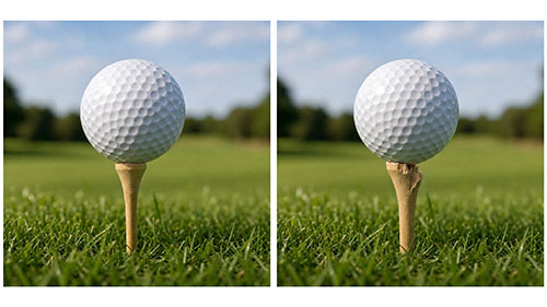
Caption: Why bone loss changes the operation. Left: a golf ball on an intact tee - the shallow socket of a normal shoulder, which holds the ball securely because the whole rim is present. Right: the same ball on a tee with a piece broken away. The ball no longer sits centered, and very little displaces it. This is what significant glenoid bone loss does, and it is why reattaching the torn tissue is not enough on its own.
The Glenoid Track
Modern management is built around the glenoid track, which explains why two similar-sounding injuries need different operations.
As the arm elevates and rotates outward, the humeral head contacts the glenoid along a predictable path - the track.
- On-track: the Hill-Sachs lesion stays within that path. The shoulder remains stable
- Off-track: the lesion extends beyond the edge, where it can engage the rim and lever the shoulder out
- Near-track: technically within the track, but close to the edge with little margin. Classified on-track, and behaves less predictably than that label suggests
Two things determine which it is: the size and position of the Hill-Sachs lesion, and how much glenoid bone has been lost - because bone loss narrows the track itself. The two lesions are two halves of one problem and are assessed together.
What the numbers mean. Twenty percent glenoid bone loss was long treated as the “critical” threshold. Outcomes begin to deteriorate well below it: “subcritical” loss of roughly 13.5 to 20 percent is associated with meaningfully worse function after soft-tissue repair alone. I use both together - the percentage tells you how much socket is gone, the track tells you whether what remains can contain the head through the range that patient uses.
After a First Dislocation: What Are the Odds?
Recurrence depends most on age, then activity level and bone loss.
| Recurrence without surgery | |
|---|---|
| Under 20 | Commonly above 70 percent; some series report 80 percent or higher in contact athletes |
| 20 - 30 | High, though lower than adolescents |
| Over 40 | Substantially lower - but a higher likelihood of an associated rotator cuff tear |
Contact and collision athletes carry higher risk within every age band, and any glenoid bone loss raises it further.
Age is the strongest single predictor, for two reasons.
- Younger tissue is more elastic. Collagen stiffens with age, so a younger shoulder has more inherent give in the capsule and ligaments. That laxity is normal and part of what allows a young athlete to move as they do - but it means the restraints are more easily stretched once the labrum has detached
- Younger patients return to the activities that caused the dislocation, often for several more seasons, and that exposure concentrates in exactly the age group where the figures are highest
For a young contact athlete, non-surgical management means accepting a high probability of recurrence rather than choosing an equivalent alternative. That may still be right for a particular person and season, but it should be chosen with the figure in view.
Diagnosis
The history does most of the work. How did it happen? Did it need to be put back in, or did it go back on its own? How many times, and with less force each time? Which position produces apprehension?
A dislocation during sleep is particularly concerning. Asleep, the muscles are at their most relaxed and are not holding the joint, and there is no sense of where the arm is in space. A shoulder that comes out under those conditions is doing so with essentially no force and no muscular defense - which indicates the passive restraints are no longer doing their job.
The examination covers:
- Apprehension and relocation testing, and load-and-shift assessment of translation
- Generalized ligamentous laxity. Widespread laxity points toward multidirectional instability rather than traumatic anterior instability
- Scapular mechanics, frequently abnormal in unstable shoulders - sometimes contributing, sometimes a consequence, and part of the rehabilitation either way
Imaging:
- X-rays confirm the joint is reduced and can show a bony Bankart fragment or Hill-Sachs lesion
- MRI defines the labral tear, capsular injury, and associated damage. A standard MRI without contrast is usually sufficient in my practice, particularly on a 3 Tesla scanner. MR arthrography is used routinely at some centers; it is not our default and is most useful where a standard scan has not answered the question
- CT is obtained selectively rather than routinely. If there is concern about the amount of bone loss, or about its location on the glenoid or humeral head side, a three-dimensional CT scan is a useful study to quantify the loss and to aid in preoperative planning. Three-dimensional CT with the humeral head digitally subtracted gives a face-on view of the socket and is the reference standard for that measurement
What Happens Next
Treatment is covered in detail on a separate page: Treating Anterior Shoulder Instability - non-surgical management, the in-season athlete, the five operations and how one is chosen, recovery, and return to sport.
Frequently Asked Questions
Will my shoulder dislocate again after the first time?
It depends heavily on age and activity. Athletes under 20 treated without surgery have recurrence commonly reported above 70 percent, and some series report 80 percent or higher in contact athletes. Risk declines with age. Patients over 40 have a much lower recurrence rate but a higher chance of an associated rotator cuff tear.
My shoulder feels loose but has never fully dislocated. Does that count?
Yes. Subluxations - partial slips that reduce on their own - can cause the same structural damage as a full dislocation. A sudden dead feeling in the arm, or a sense of the joint shifting and resetting, should be assessed.
How much glenoid bone loss is too much for an arthroscopic repair?
There is no single agreed threshold. Twenty percent was long treated as the critical figure, but outcomes deteriorate below it, and loss of roughly 13.5 to 20 percent - sometimes called subcritical - is associated with worse results after soft-tissue repair alone. Current practice weighs the percentage together with the glenoid track. If there is concern about the amount or location of bone loss, a three-dimensional CT scan quantifies it and aids preoperative planning.
My MRI report says the labral tear is from 3 to 6 o’clock. What does that mean?
Labral tears are located by position on a clock face, with the socket viewed face-on, 12:00 at the top and 6:00 at the bottom. In a right shoulder, 3:00 is the front; in a left shoulder, the front is 9:00, because the two sides are mirror images. A tear from 3:00 to 6:00 in a right shoulder - or 9:00 to 6:00 in a left - describes a Bankart lesion at the front and lower rim, the typical pattern after a forward dislocation.
What is a Hill-Sachs lesion?
A dent in the back of the ball of the shoulder, created when it rides over the front rim of the socket during a dislocation. Small ones matter little. Larger ones can catch on the rim and cause the shoulder to dislocate again, which is what the glenoid track assesses.
Does a shoulder dislocation cause arthritis later?
It can. Post-traumatic arthritis is a recognized long-term consequence of recurrent dislocations, and can also follow older stabilization techniques that overtightened the joint. (See: Glenohumeral Osteoarthritis)
Dislocated your shoulder?
Dr. Gabriella Ode is a fellowship-trained sports medicine and shoulder surgeon at the HSS Sports Medicine Institute, head team orthopaedic surgeon for the New York Liberty, and assistant team orthopaedic surgeon for the Brooklyn Nets. She treats shoulder instability in athletes at every level. Request an appointment or request a remote second opinion.
Financial disclosure.
Dr. Ode is a paid consultant for Advita Ortho and ConMed, manufacturers of orthopaedic implants and surgical instrumentation. These relationships are reported publicly through the CMS Open Payments database. She receives no compensation for this website, and no compensation is tied to any individual patient’s treatment decision. Her complete financial disclosures are available here.
References
- Shaha JS, Cook JB, Song DJ, Rowles DJ, Bottoni CR, Shaha SH. Redefining “critical” bone loss in shoulder instability: functional outcomes worsen with “subcritical” bone loss. Am J Sports Med. 2015.
- Yamamoto N, Itoi E, Abe H, et al. Contact between the glenoid and the humeral head in abduction, external rotation, and horizontal extension: a new concept of glenoid track. J Shoulder Elbow Surg. 2007.
- Di Giacomo G, Itoi E, Burkhart SS. Evolving concept of bipolar bone loss and the Hill-Sachs lesion: from “engaging/non-engaging” lesion to “on-track/off-track” lesion. Arthroscopy. 2014.
This article is for general education and is not a substitute for individual medical advice. Please consult a physician about your specific condition.