Multidirectional Shoulder Instability: When the Shoulder Is Loose in More Than One Direction
Multidirectional instability (MDI) is symptomatic instability of the shoulder in two or more directions, most often including a downward - inferior - component. It usually develops without a single injury to point to.
The presentation covers a wide range. Some patients describe only vague shoulder discomfort, reduced strength, or a decline in athletic performance. Others experience frequent subluxations - partial slips of the joint - during ordinary movements, along with apprehension, pain, and real limitation in daily activities.
MDI is a clinical diagnosis. It is made from the history and the physical examination, not from imaging.
It is most commonly seen in young female patients, typically from adolescence into the mid-thirties.
How MDI Differs From a Dislocated Shoulder
Two long-standing acronyms describe the two ends of the instability spectrum, and they remain a useful shorthand.
| TUBS | AMBRI | |
|---|---|---|
| Cause | Traumatic - a specific injury | Atraumatic - no single event |
| Direction | Unidirectional, usually anterior | Multidirectional |
| Sides | One shoulder | Bilateral - often both |
| Structure | Bankart lesion - labrum torn off the socket | Capsular laxity and redundancy |
| First-line treatment | Surgery is frequently required | Rehabilitation |
| If surgery is needed | Labral repair | Inferior capsular shift |
These are two different problems requiring two different approaches, which is why distinguishing them matters. (See: Anterior Shoulder Instability)
Why the Shoulder Becomes Unstable
The shoulder is held stable by two sets of structures working together.
Static stabilizers hold the joint passively:
- The glenohumeral ligaments
- The glenoid - the socket - and its labrum, the rim of cartilage that deepens it
- Negative pressure inside the joint, which creates a mild suction effect
Dynamic stabilizers hold the joint actively, through muscle:
- The rotator cuff
- The periscapular muscles that position the shoulder blade
- The three heads of the deltoid
- The long head of the biceps tendon
In MDI, instability results from dysfunction or imbalance in one or both systems. Patients with MDI frequently show excessive capsular laxity, capsular redundancy, an increased volume inside the joint, and deficiencies of the rotator interval - the gap at the front of the shoulder between two of the cuff tendons, which normally helps hold the joint snug.
These features are generally attributed to either congenital factors, including connective tissue disorders, or repetitive microtrauma, particularly in overhead athletes.
Hypermobility Is Not the Same as Instability
This distinction matters, and it is a common source of unnecessary concern.
Generalized joint hypermobility - being “double-jointed” - is common, particularly in adolescents, and it is common in female athletes. It frequently causes no problems whatsoever. It is a risk factor for developing MDI. It is not the diagnosis.
Symptoms are required for the diagnosis. A hypermobile shoulder that translates a long way on examination but causes no pain, apprehension, or functional limitation does not meet the criteria for MDI.
What a lax shoulder does mean is that the muscles have more work to do. Where the ligaments and capsule are loose, the neuromuscular control and the dynamic stabilizers have to work harder to keep the head of the shoulder centered in the socket. In a well-conditioned athlete that system copes, and the shoulder is asymptomatic.
The consequence is that a lax shoulder is more sensitive to fatigue and to injury. When the shoulder is overworked, or when something is injured, the muscles can no longer take up the slack, and pain and symptoms follow more readily than they would in a tighter shoulder. This is the usual explanation for why a shoulder that has been hypermobile and comfortable for years starts to hurt.
Symptoms
The characteristic complaint is vague, activity-related shoulder pain that develops gradually.
Instability is often not the primary complaint. Many patients describe nonspecific pain across the whole shoulder, or pain around the shoulder blade, with no history of injury. A decline in strength or in athletic performance - particularly in overhead sport - is a frequent early sign and is often what finally brings someone in.
Although MDI involves instability in more than one direction, most patients can identify a predominant one. Where the symptoms appear is informative:
| Direction | When symptoms occur |
|---|---|
| Inferior (the hallmark of MDI) | Carrying heavy loads below shoulder level - a backpack, a suitcase, grocery bags |
| Posterior | Removing a shirt, driving, pushing a heavy door |
| Anterior | The arm out to the side and rotated back - throwing, or reaching behind the back |
Other common features:
- A sense of the shoulder slipping, sliding, or partially coming out
- Fatigue and weakness with overhead activity
- Symptoms in both shoulders - bilateral involvement is common, and helps distinguish MDI from traumatic instability
- Occasional numbness or tingling from traction on the nerves
- No memorable injury - most patients cannot identify a specific event
- The ability to shift the shoulder out of position deliberately in some patients, which is important to mention to your surgeon, as it changes the treatment discussion
A medical and family history of multiple joint dislocations, or of features suggesting a connective tissue disorder, raises suspicion for MDI.
MDI in Women Athletes
Hypermobility is common in female athletes, and on its own it is not a problem. In many sports it is an advantage. A hypermobile shoulder can be entirely comfortable and perform at a high level for years.
What a lax shoulder is, is less forgiving. Because the passive restraints are loose, the dynamic stabilizers and the neuromuscular control system carry more of the load. What I see clinically is that this makes the shoulder more sensitive - to fatigue when it is being overworked, and to pain when something in it has been injured. The same training error or minor injury that a tighter shoulder absorbs without complaint will produce symptoms in a lax one.
Pain is what brings patients in, not instability. Very few describe the shoulder as unstable. They describe it as aching, tired, or unreliable. Because the complaint is pain, tendinitis is a reasonable initial working diagnosis, and it is the examination - laxity in more than one direction, with the patient’s own symptoms reproduced - that reframes it.
Symptoms are frequently present on both sides, which tends to normalize them. A shoulder that has always felt this way, and a second shoulder that feels the same, does not register as a problem to be reported.
Hypermobility is often an asset in the sport. In swimming, gymnastics, and dance, range of motion is trained for and rewarded. Patients are unlikely to volunteer it as a symptom, and may be reluctant to see it as connected to the pain.
The trigger is usually a change rather than an injury - a growth spurt, a jump in training volume, a return from time off, or the transition between seasons.
The prognosis is good, and it should be said early. Most athletes with MDI return to their sport. The treatment asks for patience rather than surgery, and the conversation goes better when that is established at the first visit rather than after a few months of frustration.
Sport by Sport

Caption: Swimmer’s shoulder. Symptoms characteristically build through a training session, as the muscles holding the shoulder stable fatigue, and settle with rest - which is what distinguishes them from the pain of a structural injury, and why they are easy to attribute to ordinary training soreness.
Swimming - “swimmer’s shoulder.” The highest overhead volume of any sport, with thousands of shoulder revolutions per session. The sport also selects for laxity, since range of motion makes for an efficient stroke. Freestyle and butterfly repeatedly load the front and underside of the capsule. Symptoms characteristically appear late in a session, as the dynamic stabilizers fatigue, and settle with rest - which is why they are easy to attribute to ordinary training soreness. Training volume, paddle use, stroke mechanics, and one-sided breathing all modify it.
Stroke matters. Some studies find butterfly the most likely to provoke pain, because of its repeated internal and external rotation. Breaststroke generally places the least stress on the shoulder, since it does not require the arms to be raised overhead or out to the side as much as the other strokes.
Conditioning can usually be maintained through a painful period - kicking with the arms at the side, then working up through lower-stress strokes, with shorter repetitions and distances before returning to a normal routine. Swimming through pain tends to produce a cycle of recurrence rather than a return to form.
I discussed shoulder pain in swimmers, including multidirectional instability and rotator cuff tendinitis, in an article for U.S. Masters Swimming.
Throwing - “thrower’s shoulder.” Years of throwing gradually increase external rotation in the dominant arm. That asymmetry is an adaptation, not a diagnosis - it is present in throwers with no symptoms at all. Distinguishing a thrower’s shoulder from true MDI matters, because the laxity is a different kind: acquired rather than constitutional, in one direction, in one arm, whereas MDI is generalized and usually affects both shoulders. Loss of scapular control frequently accompanies it. (See: Scapular Dyskinesis)
Gymnastics. Unique in making the shoulder a weight-bearing joint. The sport both selects for hypermobility and then loads that lax shoulder overhead under body weight - handstands, bar work, vaulting. Symptoms concentrate in support positions.
Dance. Extreme range of motion is trained deliberately, and partnering work adds overhead load. Ballet in particular selects strongly for generalized hypermobility, so a high Beighton score in this group is expected rather than remarkable.
Volleyball. Repeated maximum external rotation in the arm cock, deceleration through the arm swing, and blocking with both arms overhead.
Basketball. Less classically associated, but overhead demand combined with contact means both traumatic and atraumatic patterns are seen.
Weight training. Repetitive pressing with the arms forward and near the midline - bench press and push-up patterns - drives the head of the shoulder backward and loads the back of the joint. This produces a separate, one-directional problem rather than MDI. (See: Posterior Labral Tears and Posterior Instability)
Terms You May Hear
Several terms describe conditions along the shoulder laxity spectrum. They overlap, and none of them is interchangeable with MDI.
| Term | What it means |
|---|---|
| Swimmer’s shoulder | A descriptive umbrella term for shoulder pain in swimmers, not a single diagnosis. Historically used interchangeably with impingement; more often reflects fatigue of the dynamic stabilizers, cumulative tendinopathy, and loss of scapular control. |
| Thrower’s shoulder | The pattern of adaptation and injury seen in the throwing arm, including acquired laxity in the dominant shoulder. |
| SICK scapula | A described pattern of scapular malposition and dysfunction in overhead athletes. (See: Scapular Dyskinesis) |
| Benign joint hypermobility | Hypermobility without symptoms. Common, and not a diagnosis requiring treatment. |
| AMBRI | The classic shorthand for the MDI pattern - atraumatic, multidirectional, bilateral, rehabilitation, inferior capsular shift. |
| Capsular redundancy / patulous capsule | Terms used on MRI reports for a capsule that is loose and larger in volume than normal. |
| Microinstability | Translation of the head of the shoulder too small to be felt as a slip, but enough to produce symptoms. |
MDI and Posterior Instability
These overlap, and the distinction affects treatment.
Posterior instability can be its own problem - from trauma, or acquired through repetitive loading of the arm forward and near the midline, as in bench press and push-up patterns. In that setting the shoulder is unstable in one direction, and there may be structural damage to the posterior labrum or early wear on the back of the socket.
In MDI, posterior symptoms are one component of instability in several directions, in a shoulder that is globally lax, usually bilateral, and usually without structural injury.
Both can produce posterior symptoms, so both belong in the examination. Where posterior symptoms dominate, or where a specific loading pattern explains them, the posterior instability pathway is the more relevant one. (See: Posterior Labral Tears and Posterior Instability)
How It Is Diagnosed
MDI is diagnosed from the history and the physical examination.
The Examination
Range of motion is assessed both actively and passively, upright and then lying down so the shoulder blade is properly stabilized, and always compared with the other side.
Shoulder-specific hypermobility is suggested by:
- Passive external rotation greater than 85 degrees with the arm at the side
- Passive abduction greater than 110 degrees - the Gagey test
Generalized hypermobility is assessed with the Beighton score, a nine-point scale covering the elbows, knees, thumbs, and little fingers on both sides, plus forward bending. A score of 4 or more indicates generalized joint hypermobility.
Instability testing in multiple directions:
- Sulcus sign - with the arm relaxed at the side, downward traction produces a visible dimple below the point of the shoulder. Greater than 2 cm is considered positive, and this is the classic finding in MDI. A sulcus that persists when the arm is rotated outward points to slack in the rotator interval
- Anterior and posterior drawer tests
- Load and shift, apprehension and relocation, jerk test, and Gagey
One point matters more than any of the individual tests: reproducing the patient’s own symptoms - their pain, their apprehension - carries more clinical weight than how far the head of the shoulder translates, or how high the Beighton score is.
Scapular mechanics. Scapular dyskinesis frequently contributes to MDI and is assessed during active motion. Reduced upward rotation, winging of the inner border of the shoulder blade, and forward tilting during elevation are the signs to look for.
Strength, tested on both sides in several planes and functional positions - rotator cuff, scapular stabilizers, deltoid, periscapular muscles, and core - to identify the imbalances driving the loss of control.
Posture, including spinal alignment and the resting position of the shoulder blade. Forward head posture, rounded shoulders, and a protracted shoulder blade all alter dynamic stability.
The neck and mid-back. Cervical and thoracic mobility and strength are assessed to identify restrictions or movement patterns contributing to the picture. (See: Shoulder Pain vs. Cervical Radiculopathy)
Imaging
MDI is not diagnosed on imaging. The findings associated with it - a loose, roomy capsule, for example - are also seen in people whose shoulders are entirely comfortable, so the scan is interpreted alongside the history and the examination rather than on its own. Imaging is used to assess the bone, to identify associated injury, and to exclude other problems.
X-rays are usually normal in MDI, but a complete series is important. It evaluates the alignment of the ball within the socket and identifies structural contributors - glenoid dysplasia, a bony Bankart lesion, a Hill-Sachs lesion, or bone loss. The standard views:
| View | What it shows |
|---|---|
| True AP (Grashey) | The joint space and how well the ball is centered in the socket |
| AP in internal and external rotation | The contour of the humeral head and any Hill-Sachs lesion |
| Scapular Y | Dislocation or subluxation, and the orientation of the shoulder blade |
| Axillary | Critical. Subtle subluxation, alignment, and bone loss |
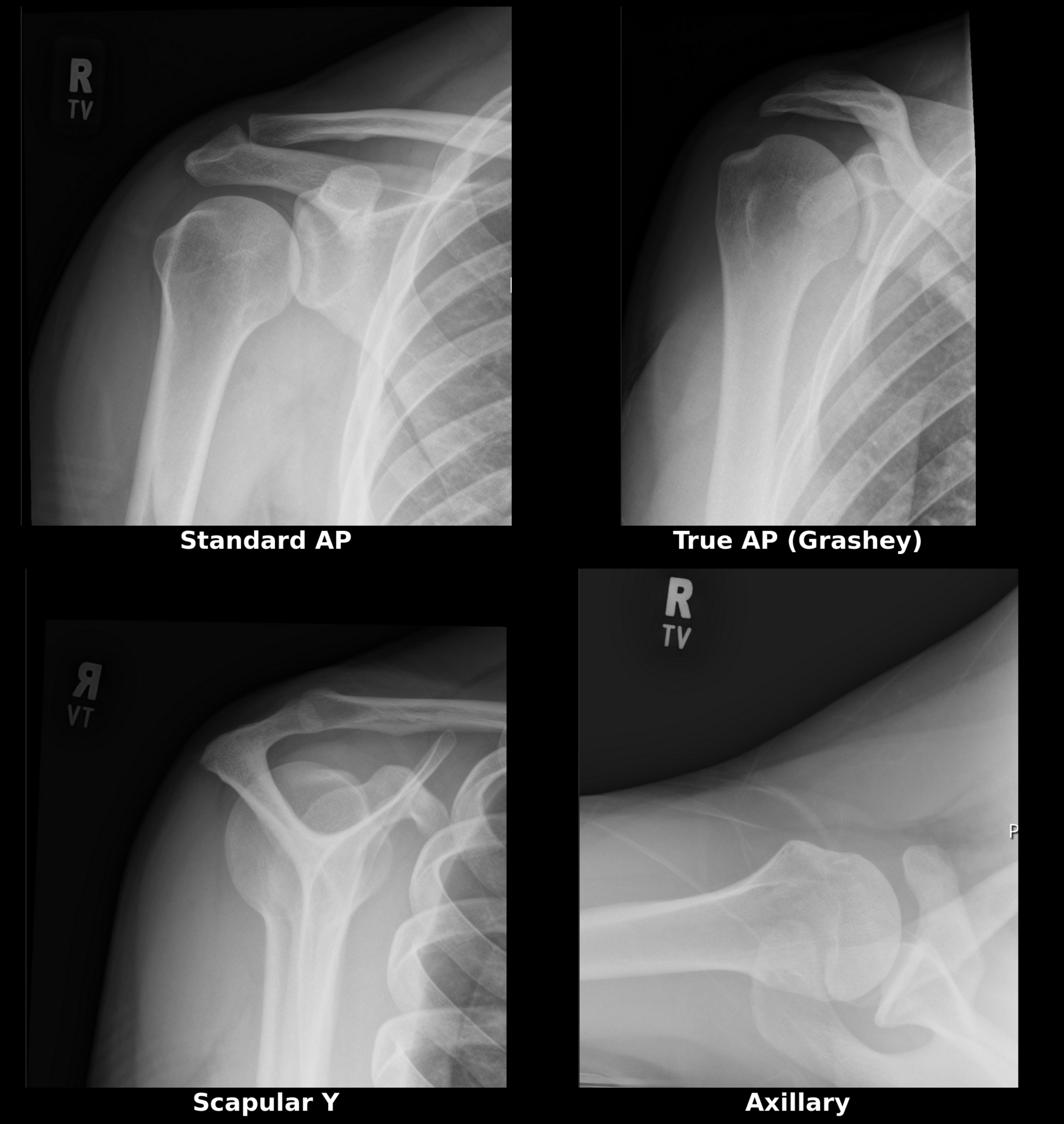
Caption: A complete shoulder X-ray series, normal throughout. The four standard views: a standard AP, a true AP (Grashey), a scapular Y, and an axillary. In multidirectional instability the X-rays are typically normal, as they are here. The series is obtained to assess the bone, to identify structural contributors such as glenoid dysplasia or bone loss, and to exclude other causes of the symptoms - not to make the diagnosis, which is made from the history and the examination.
MRI. A high-resolution 3 Tesla MRI gives excellent detail of the capsule and the labrum. At some centers, an MRI with dye - an arthrogram - is performed to provide greater detail of the joint space.
Findings that support the diagnosis include capsular redundancy, an abnormally wide inferior capsule, increased volume within the joint, and widening of the rotator interval. MRI is also useful for identifying associated damage - a Bankart lesion, a Kim lesion of the posterior labrum, or a chondrolabral injury.
There is no single universally accepted set of MRI criteria for MDI. Several measurements have been proposed and studied, but none has been adopted as definitive - which is another reason the diagnosis rests on the examination.
When to Look Beyond the Shoulder
Some patients with MDI have a generalized connective tissue disorder, and the shoulder is one manifestation of it. Referral for further evaluation is appropriate where there is symptomatic hypermobility in several joints, or other features suggesting a collagen disorder - including cardiac conditions or postural orthostatic tachycardia syndrome.
Features in the history that raise this possibility:
- Joint dislocations or subluxations, particularly at several sites
- Easy bruising or bleeding
- Poor wound healing
- Family history
Features on examination, largely of the skin: abnormal or stretched scars, brown discoloration from repeated bruising, striae, small firm nodules under the skin, and skin that stretches easily when pinched at the forearm.
A cardiac examination is performed in these patients to assess for valve or vessel abnormalities.
Hypermobile Ehlers-Danlos syndrome carries a substantial musculoskeletal burden, and chronic pain is reported near-universally. Identifying it changes the expectations set around treatment, including around surgery, and brings in the specialists who should be managing the broader condition.
Treatment: Rehabilitation First
A comprehensive, structured rehabilitation program is the first-line treatment for MDI. Its purpose is to restore the function of the dynamic stabilizers so that the laxity of the passive structures stops producing symptoms.
Exercise cannot tighten a lax capsule. What it can do is train the muscles that control the joint to hold the head of the shoulder centered in the socket - restoring what is called the concavity-compression mechanism, by which the cuff actively presses the ball into the socket.
A program generally proceeds in stages:
- Education, so the patient understands the anatomy, the role of the dynamic stabilizers in their symptoms, and what the program is doing. This is not preamble - engagement is closely tied to whether the program works
- Activity modification, avoiding the positions that provoke symptoms during the early phase. Taping may help support the shoulder
- Motor control work, correcting the patient’s specific scapular control deficits and training the head of the shoulder to stay centered
- Strengthening of the rotator cuff and periscapular muscles, with attention to movement quality rather than load alone
- Progression to functional and sport-specific movement
Supervised, structured, phase-specific programs have been shown to improve scapular upward rotation, muscle strength, movement quality, and patient-reported function in atraumatic shoulder instability.
In the absence of significant structural injury - a Bankart lesion or a bony defect - a non-operative program should be pursued for at least six months before surgical options are considered. That is the figure in the published guidance. The interval is not fixed, and it is often extended - sometimes to a year - where a patient is continuing to make gains, or where therapy has been interrupted and the program has not had a fair run. Swimmers and throwing athletes commonly fall into that group.
Improvement is gradual rather than sudden, and the timeline should be clear at the outset. What that period describes is a program worked all the way through - progressing through each stage of therapy to its goals, with the home exercises done consistently alongside the supervised sessions. Both halves matter. The home program is where most of the repetition happens, and the gains in motor control come from that repetition.
The Maintenance Program
Formal therapy ends. The exercises should not.
Even after a full course of therapy and a good result, patients are counseled to continue a maintenance program. The shoulder is still lax - that does not change - so the strength and motor control built during rehabilitation are what keep it comfortable. When the exercises stop, that capacity gradually falls away, and symptoms can return with the next increase in training or the next period of heavy demand.
What maintenance looks like is a short, regular routine rather than a continuation of full rehabilitation: rotator cuff and scapular work a few times a week, folded into an existing training program or done at home.
Scapular control is the part most commonly carried forward. The muscles that position the shoulder blade set the socket underneath the arm, and in a lax shoulder that positioning does more of the work than it does in a tight one. My Scapular Mechanics Program is a printable home program built for exactly this, and it works well as an ongoing maintenance routine once formal therapy has finished. (The condition it was written for is explained here: Scapular Dyskinesis.)
For broader general conditioning, the AAOS shoulder conditioning program is a reasonable additional resource.
Think of it as ongoing shoulder wellness in the same way an athlete maintains core strength or flexibility - not as treatment that never finishes.
When Surgery Is Considered
Surgery is considered for patients with persistent, disabling instability after a diligent course of therapy. Published guidance generally puts that at longer than six to nine months.
In practice the interval varies. Where a patient is still making gains, or where therapy was interrupted and the program has not had a continuous run, the trial is often extended - sometimes to a year. Swimmers and throwing athletes frequently fall into this category, since their sport both selected for the laxity and then loaded it, and they tend to respond over a longer timeframe than other patients with MDI.
The operation addresses the static stabilizers. The standard procedure is a capsular shift, performed either open or arthroscopically, in which the redundant capsule is overlapped and tightened, reducing the volume within the joint. The plication is centered on the inferior glenohumeral ligaments. Where the capsule is lax throughout, an arthroscopic pancapsular shift addresses it circumferentially. If the rotator interval is slack, it may be closed at the same time.
Reported outcomes are good. Series of arthroscopic pancapsular shift report significantly improved patient-reported outcome scores and low rates of recurrent instability, including at a mean follow-up of nine years.
Outcomes do vary, and two points belong in the conversation:
- In one series of athletes, female patients with atraumatic MDI had lower postoperative outcome scores, higher rates of persistent subluxation, and lower rates of return to sport than other groups. This is not a reason to withhold surgery from a patient who needs it. It is a reason to be thorough about the rehabilitation program first, and specific about expectations
- In patients with a diagnosed connective tissue disorder, expectations are adjusted accordingly, and recurrence rates are higher
Rehabilitation after surgery is prolonged, and the same neuromuscular retraining is required afterward. The operation changes the passive restraint; it does not supply the motor control.
This variability in outcome is why surgery is generally reserved for cases that have not responded to an extensive trial of therapy - the sequence is part of the treatment, not a preliminary to it.
Frequently Asked Questions
What is multidirectional shoulder instability?
It is symptomatic instability of the shoulder in two or more directions, usually including a downward component, and usually occurring without a significant injury. It generally results from a naturally lax joint capsule - sometimes worsened by repetitive overhead activity - combined with insufficient muscular control of the joint.
How is MDI different from a dislocated shoulder?
A traumatic dislocation involves a specific injury that tears the labrum off the socket, and it is usually in one direction. MDI typically has no injury, involves several directions, is often present in both shoulders, and is associated with generalized ligamentous laxity. Traumatic instability frequently requires surgery; MDI is treated first with rehabilitation.
Can multidirectional instability be treated without surgery?
Yes, and it usually should be. A structured program targeting rotator cuff strength, scapular control, and neuromuscular retraining is the first-line treatment. Where there is no significant structural injury, published guidance recommends a trial of at least six months before surgical options are considered. In practice the trial is often extended - sometimes to a year - where a patient is still improving, or where therapy has been interrupted. Swimmers and throwing athletes frequently fall into that group.
Is being “double-jointed” the same as having shoulder instability?
No. Generalized joint hypermobility is common, is particularly common in female athletes, and frequently causes no problems. A Beighton score of 4 or more indicates generalized hypermobility, but hypermobility alone is not a diagnosis. It becomes a clinical issue only when it produces symptoms - pain, apprehension, or functional limitation.
If my shoulder is loose but does not hurt, why did it start hurting now?
A lax shoulder relies more heavily on its muscles. The ligaments and capsule provide less passive support, so the rotator cuff, the muscles controlling the shoulder blade, and the neuromuscular control system have to work harder to keep the joint centered. That system usually copes well. It copes less well when the shoulder is fatigued from being overworked, or when something has been injured - which is why a hypermobile shoulder that has been comfortable for years can become painful after a change in training or a minor injury.
Why is MDI more common in women?
Women have on average greater ligamentous laxity than men, influenced by both connective tissue composition and hormonal factors. MDI is also common in sports with high female participation and high overhead demand - swimming, gymnastics, dance, and volleyball - where repetitive end-range motion further loads the capsule.
What is a positive sulcus sign?
With the arm relaxed at the side, downward traction on the arm opens a visible gap below the point of the shoulder. A gap greater than 2 cm is considered positive and indicates downward laxity. It is the classic examination finding in MDI.
Do I need an MRI to diagnose MDI?
No. MDI is diagnosed from the history and the physical examination. Imaging is used to assess the bone and to identify associated injury rather than to make the diagnosis. X-rays should be a complete series including an axillary view. Where soft-tissue detail is needed, a 3 Tesla MRI provides it. At some centers, an MRI with dye - an arthrogram - is performed to give greater detail of the joint space.
How long does rehabilitation take for MDI?
Improvement is gradual. Plan on a minimum of six months, working all the way through the stages of a supervised therapy program and keeping up the home exercises alongside it - and understand that many patients keep improving well beyond that. Where gains are still coming, the program is often continued rather than cut short at a fixed date. Even after a good result, patients are advised to continue a maintenance program long-term. The shoulder remains lax, so the strength and motor control built during rehabilitation are what keep it comfortable. The Scapular Mechanics Program works well for this. Consistency matters more than intensity.
Will I need to stop my sport?
Usually not permanently. Most athletes with MDI return to their sport, often with temporary modification of training volume during the initial rehabilitation phase and an ongoing maintenance program afterward.
Shoulder feels loose or unreliable?
Dr. Gabriella Ode is a fellowship-trained sports medicine and shoulder surgeon at the HSS Sports Medicine Institute with a practice focus on the care of women athletes of all ages. Request an appointment or request a remote second opinion.
Financial disclosure.
Dr. Ode is a paid consultant for Advita Ortho and ConMed, manufacturers of orthopaedic implants and surgical instrumentation. These relationships are reported publicly through the CMS Open Payments database. She receives no compensation for this website, and no compensation is tied to any individual patient’s treatment decision. Her complete financial disclosures are available here.
This article is for general education and is not a substitute for individual medical advice. Please consult a physician about your specific condition.
References
- Johnson DJ, Tadi P. Multidirectional shoulder instability. StatPearls. 2023.
- Housset V, Ho SWL, Lädermann A, Phua SKA, Hui SJ, Nourissat G. Multidirectional instability of the shoulder: a systematic review with a novel classification. EFORT Open Rev. 2024.
- Merolla G, Cerciello S, Chillemi C, Paladini P, De Santis E, Porcellini G. Multidirectional instability of the shoulder: biomechanics, clinical presentation, and treatment strategies. Eur J Orthop Surg Traumatol. 2015.
- Watson L, Pizzari T, Balster S, Lenssen R, Warby SA. Advances in the non-operative management of multidirectional instability of the glenohumeral joint. J Clin Med. 2022.
- Watson L, Warby S, Balster S, Lenssen R, Pizzari T. The treatment of multidirectional instability of the shoulder with a rehabilitation programme: Parts 1 and 2. Shoulder Elbow. 2016 and 2017.
- Emery R, Mullaji A. Glenohumeral joint instability in normal adolescents: incidence and significance. J Bone Joint Surg Br. 1991.
- Fithian AT, Edmonds EW, Egger AC, et al. Evaluation and management of glenohumeral instability in adolescent patients: an expert consensus statement. Orthop J Sports Med. 2024.
- Kay J, Heyworth BE, Milewski MD, Kramer DE. Pediatric and adolescent shoulder instability. Curr Rev Musculoskelet Med. 2023.
- Gruskay JA, Rakowski DR, Woolson TE, Horan MP, Millett PJ. Clinical outcomes after arthroscopic pancapsular shift for the treatment of multidirectional glenohumeral instability at a mean follow-up of 9 years. Am J Sports Med. 2022.
- Raynor MB, Horan MP, Greenspoon JA, Katthagen JC, Millett PJ. Outcomes after arthroscopic pancapsular capsulorrhaphy with suture anchors for the treatment of multidirectional glenohumeral instability in athletes. Am J Sports Med. 2016.
- Diep D, Gemae MR, Farag J, Tay MRJ, Mohankumar R, Mittal N. Imaging modalities for atraumatic shoulder hypermobility: a scoping review. Skeletal Radiol. 2024.
- Wanivenhaus F, Fox AJS, Chaudhury S, Rodeo SA. Epidemiology of injuries and prevention strategies in competitive swimmers. Sports Health. 2012.
- Burkhart SS, Morgan CD, Kibler WB. The disabled throwing shoulder: spectrum of pathology. Part I: pathoanatomy and biomechanics. Arthroscopy. 2003.
- Kibler WB, Kuhn JE, Wilk K, et al. The disabled throwing shoulder: spectrum of pathology - 10-year update. Arthroscopy. 2013.