Cuff Tear Arthropathy: When a rotator cuff tears Becomes Arthritis
Rotator cuff tear arthropathy is a specific form of shoulder arthritis caused by a large, long-standing rotator cuff tears. When the rotator cuff has been torn for years, the ball of the shoulder drifts upward out of its socket and begins grinding against the underside of the acromion - the bony roof of the shoulder. Over time this produces a distinctive pattern of joint destruction that looks and behaves differently from ordinary shoulder arthritis.
It is worth understanding as its own condition rather than as “arthritis with a cuff tear,” because the mechanics are different, the symptoms are different, and - critically - the treatment is different. An operation that works well for ordinary shoulder arthritis will fail in a shoulder with cuff tear arthropathy.
How it develops
The rotator cuff does two jobs. The obvious one is powering rotation. The less obvious one, and the one that matters here, is holding the ball centered in the socket so the deltoid can lift the arm efficiently.
When a massive rotator cuff tears goes unrepaired for years, that centering force disappears. A predictable sequence follows:
- Superior migration. The unopposed pull of the deltoid drags the humeral head upward. On X-ray, the space between the humeral head and the acromion narrows and eventually disappears - one of the defining radiographic findings.
- Acetabularization. The undersurface of the acromion, which was never designed to bear load, is repeatedly ground against by the humeral head. It gradually erodes into a smooth, cup-shaped surface, and the two bones effectively form a false joint.
- Femoralization. Meanwhile, the humeral head itself loses its normal round contour, becoming rounded off and smoothed at the top.
- Cartilage loss and joint destruction. The cartilage of the true glenohumeral joint wears away, and the bone can soften and collapse.
This process typically unfolds over many years, which is why cuff tear arthropathy is predominantly a condition of patients in their seventies and eighties, and why it is more common in women.
Not every massive rotator cuff tears leads to cuff tear arthropathy. Many people live for decades with large tears and never develop it. Why some shoulders progress and others don’t isn’t fully understood - biology, inflammation, and individual anatomy all appear to contribute.
Symptoms
The hallmark combination is pain plus a striking loss of the ability to lift the arm, in a patient who often cannot recall a specific injury.
What patients typically describe:
- Deep aching pain, often severe at night, frequently the reason they finally seek care
- Weakness that has crept up over years rather than appearing suddenly
- Inability to raise the arm overhead - some patients cannot get the arm past shoulder height, and some cannot lift it away from their side at all
- A “shrugging” motion when attempting to raise the arm, as the shoulder blade compensates for a shoulder that can’t
- Grinding or clunking with movement
- Difficulty with basic tasks - reaching a shelf, washing hair, putting on a coat
Pseudoparalysis is the term for the most severe version: the deltoid is strong, the nerves are intact, but the arm cannot be actively lifted because the mechanics have failed. Patients can often hold the arm up if someone else lifts it there, which is a useful examination finding and frequently the moment the diagnosis becomes clear.
How it’s diagnosed
Cuff tear arthropathy is usually diagnosed on physical examination and plain X-rays.
Examination distinguishes active from passive motion - the gap between them is the key finding. It also tests the individual rotator cuff tendons, assesses deltoid function, and rules out nerve causes of weakness. Weakness from a pinched nerve in the neck can mimic this picture and must be excluded.
X-rays show the characteristic findings: loss of the acromiohumeral interval, superior migration of the humeral head, acetabularization of the acromion, and glenohumeral joint space loss.
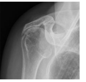
Caption: Pre-operative X-ray in cuff tear arthropathy. The humeral head has migrated upward until it sits directly against the underside of the acromion - the space that should separate them is gone.
This is what the condition looks like, and it is usually recognizable at a glance. In a healthy shoulder there is a clear gap between the top of the humeral head and the acromion above it, held open by the rotator cuff. Here that gap has disappeared entirely, because the tendon that was holding the ball down is no longer there to do it.
Which X-ray views are taken matters here more than almost anywhere else in shoulder care, because the central measurement - the acromiohumeral interval, the space between the top of the humeral head and the underside of the acromion - is easily distorted by how the film is positioned.
Two views are essential:
- The true AP view (Grashey view). A standard AP shoulder X-ray is taken straight on relative to the body, so the ball and socket overlap on the image and the joint space is partly hidden. Because the shoulder blade sits at an angle on the ribcage, a true AP is angled to match that plane - roughly 30 to 45 degrees oblique - so the beam passes cleanly through the joint. This is the view that shows the acromiohumeral interval and the glenohumeral joint space accurately rather than approximately.
- The axillary view, taken from below with the arm slightly elevated. This shows the relationship between ball and socket from underneath and defines the pattern of glenoid wear - information that directly shapes the surgical plan.
These views are not always obtained in an emergency room or primary care setting, where a single standard AP is often all that’s taken. That’s reasonable for screening for fracture or dislocation, but it is not sufficient for characterizing cuff tear arthropathy. A narrowed acromiohumeral interval can be missed or exaggerated on a poorly positioned film.
These are two views I almost always obtain when evaluating a new patient with shoulder pain. Bring any outside imaging to your appointment - it’s always useful - but don’t be surprised if the X-rays are repeated, because with this condition the views are what make the findings measurable rather than impressionistic.
MRI confirms the extent of the tear, whether it is repairable, and - importantly - the degree of fatty infiltration and atrophy in the cuff muscles. Muscle that has been retracted and fatty-infiltrated for years will not recover function even if the tendon could be reattached. This finding often settles the question of whether repair is realistic.
CT scan is obtained for surgical planning, to define glenoid bone loss and generate the three-dimensional plan used for navigated arthroplasty.
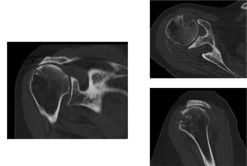
Caption: Pre-operative CT of the same shoulder. The scan shows the pattern and extent of glenoid wear, and how much bone remains to anchor an implant against - detail the X-ray cannot provide.
In a shoulder like this one, the CT is doing something specific: it tells me how much bone is left to work with. That determines where the baseplate can be anchored and how the screws must be angled to find solid bone - decisions I would otherwise be estimating in the operating room.
Treatment
Non-surgical treatment is appropriate first-line care and is sufficient for some patients, particularly those whose pain is manageable and who have adapted their activities. It includes activity modification, physical therapy focused on deltoid and scapular strengthening (deltoid-based rehabilitation programs can meaningfully improve function in selected patients), anti-inflammatory medication, and corticosteroid injections. What it cannot do is reverse the structural changes.
Rotator cuff repair is generally not an option in established cuff tear arthropathy. By this stage the tendon is retracted, the muscle is fatty-infiltrated, and repair attempts have high failure rates. Repair addresses neither the arthritis nor the altered mechanics.
Reverse total shoulder replacement is the definitive treatment, and cuff tear arthropathy is the exact problem the operation was designed to solve. By switching the ball and socket, the reverse design allows the deltoid to lift the arm without any contribution from the rotator cuff, while resurfacing the destroyed joint.
Outcomes in this population are among the most rewarding in shoulder surgery. Patients who could not lift their arm frequently regain functional overhead reach, and the improvement in night pain is usually rapid and dramatic. Published implant survivorship is approximately 91 - 98% at five years and roughly 88 - 90% at ten - figures that are particularly favorable given that most of these patients are in their seventies or eighties at the time of surgery.
An anatomic total shoulder replacement is contraindicated here. Without a functioning rotator cuff, the ball will ride upward against the socket component and cause early loosening - the so-called “rocking horse” failure. This is precisely why distinguishing cuff tear arthropathy from ordinary glenohumeral arthritis matters so much.
Why navigation is especially useful in these cases. Cuff tear arthropathy shoulders often have eroded, superiorly worn glenoid bone, which makes secure baseplate fixation more demanding. Planning screw trajectory and baseplate position on a three-dimensional CT model, and verifying that placement in real time during surgery, is most valuable exactly when bone stock is compromised.
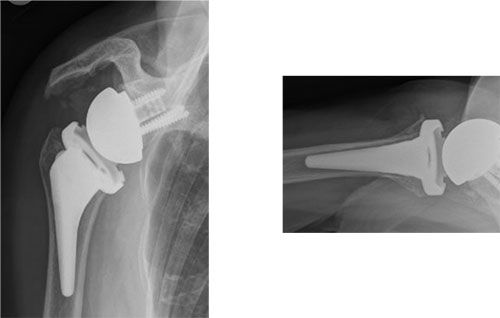
Caption: The same shoulder after reverse total shoulder replacement, shown on AP and axillary views. The ball is now fixed to the shoulder blade and the socket to the arm bone, allowing the deltoid to lift the arm without a rotator cuff.
This patient was 84 years old. That is worth stating, because age is the most common reason patients assume this operation isn’t available to them. It usually isn’t the deciding factor - overall medical health matters far more, and reverse replacement is performed routinely and successfully in patients in their eighties.
Frequently Asked Questions
What is rotator cuff tear arthropathy?
It is a specific type of shoulder arthritis that develops after a large rotator cuff tears has gone unrepaired for many years. Without the cuff to hold the ball centered, the humeral head migrates upward and grinds against the acromion, gradually destroying both surfaces along with the cartilage of the main joint.
Can cuff tear arthropathy be fixed with a rotator cuff repair?
Generally not. Once arthropathy is established, the tendon is retracted and the muscle has undergone fatty degeneration, so repair rarely restores function - and repair does nothing for the arthritis that has already developed. Reverse shoulder replacement is the treatment that reliably addresses both problems.
What X-ray views are needed to diagnose cuff tear arthropathy?
A true AP (Grashey) view and an axillary view. The key measurement is the acromiohumeral interval - the space between the humeral head and the acromion - and it can be distorted on a standard AP film taken straight on relative to the body. A true AP is angled to match the plane of the shoulder blade so the beam passes cleanly through the joint. Emergency rooms and primary care offices often obtain only a single standard AP, which is appropriate for screening for fracture but may not accurately show the findings that define this condition.
Is cuff tear arthropathy the same as regular shoulder arthritis?
No. Ordinary glenohumeral osteoarthritis is primary cartilage wear in a shoulder with an intact rotator cuff. Cuff tear arthropathy is joint destruction driven by altered mechanics after a massive cuff tear. They look different on X-ray, present differently, and require different operations.
How long does it take for a rotator cuff tears to turn into arthropathy?
Typically many years, often a decade or more, and it does not happen to everyone. Many people with large rotator cuff tears never develop arthropathy. There is currently no reliable way to predict who will.
Will I be able to lift my arm again after treatment?
Most patients who undergo Reverse shoulder replacement for cuff tear arthropathy regain the ability to raise the arm to or above shoulder height, and pain relief is typically substantial. Rotation - especially reaching behind the back - improves less predictably, which is worth discussing in detail before surgery.
I’m in my eighties. Am I too old for surgery?
Age alone is not a barrier. Reverse shoulder replacement is frequently performed successfully in patients in their eighties, and registry data actually show higher implant survivorship in older patients, since the implant faces fewer years and lower demands. What matters is your overall medical health, which your surgeon and medical team will assess together.
Living with a shoulder you can’t lift?
Dr. Gabriella Ode is a fellowship-trained shoulder surgeon at the HSS Sports Medicine Institute with advanced expertise in reverse and revision shoulder arthroplasty, performed with computer navigation. Request an appointment or request a remote second opinion.
Financial disclosure.
Dr. Ode is a paid consultant for Advita Ortho and ConMed, manufacturers of orthopaedic implants and surgical instrumentation. These relationships are reported publicly through the CMS Open Payments database. She receives no compensation for this website, and no compensation is tied to any individual patient’s treatment decision. Her complete financial disclosures are available here.
This article is for general education and is not a substitute for individual medical advice. Please consult a physician about your specific condition.