Computer-Navigated Shoulder Replacement: Why Precision Changes Outcomes
Computer navigation lets a shoulder surgeon plan an implant’s exact position on a three-dimensional model of your own anatomy before surgery, then confirm in real time during the operation that the implant is going exactly where it was planned. It is not a robot, and it does not perform any part of the surgery. It is a measurement and verification system - the surgical equivalent of GPS, which is where the technology gets its name.
I use the AdvitaGPS platform for shoulder arthroplasty, and by number of navigated cases performed I am among the top 5% of surgeons worldwide.
This page explains what navigation actually does, why the accuracy it provides matters more in the shoulder than most patients realize, and what the published evidence shows.
The problem navigation solves
The most technically demanding part of a shoulder replacement is not the ball. It is the socket.
The glenoid - the socket side of the shoulder joint, on the shoulder blade - is small, shallow, and surrounded by a limited amount of bone. By the time a patient needs a replacement, arthritis has usually eroded that socket unevenly, most often wearing away the back edge and tilting the whole surface backward. The technical term is retroversion, and it is the norm rather than the exception in an arthritic shoulder.
The surgeon’s task is to place a component squarely on that irregular, tilted, partially eroded surface, correcting the deformity where possible, seated on good bone, at the right angle in two planes, through an incision that offers a restricted view of a structure that lies deep inside the shoulder.
Why it matters that this be exact: decades of registry data identify glenoid component loosening as the leading cause of failure in anatomic total shoulder replacement, and component malposition as a major contributor. In reverse replacement, baseplate position and screw trajectory affect stability, range of motion, notching, and the security of fixation. A component placed a few degrees off is under mechanical disadvantage from the first day, and that disadvantage compounds over years.
Why it’s hard to be exact without help: plain X-rays show the glenoid in two dimensions, and version - the angle that matters most - is a three-dimensional property that X-rays systematically misrepresent. Studies of conventional freehand technique have repeatedly found meaningful variability between intended and achieved component position, even in experienced hands. This is not a criticism of surgeons; it is a consequence of asking the human eye to judge angles in a deep, obliquely oriented, partially obscured space.
How navigation works, step by step
- A preoperative CT scan. Instead of relying on X-rays, we obtain a CT of your shoulder. This produces a true three-dimensional dataset of your specific bone.
- A three-dimensional plan. The CT is converted into a model of your scapula and humerus. Before the day of surgery, I plan the operation on that model: implant type and size, the exact position of the component, the correction of version and inclination, and - for reverse replacements - the trajectory and length of each fixation screw. If a case is going to be difficult, this is where I find out, with time to solve it rather than in the operating room.
- Intraoperative registration. During surgery, a small tracker is attached to the scapula, and specific anatomic landmarks are registered so the navigation system knows precisely how your actual anatomy corresponds to the preoperative model.
- Real-time guidance. As I prepare the glenoid and place the implant, the system displays the live position and angle of each instrument relative to the plan. Instead of estimating whether the guide pin is at the intended angle, I can see the actual number.
- Verification. Before final implantation, the system confirms that the achieved position matches the plan. If it does not, it is corrected then - rather than discovered on a post-operative X-ray.
The entire system is compact, sits within the sterile field, and adds only a modest amount of time to the procedure.
Case one: a stemless replacement, start to finish
Rather than describe this abstractly, here is a single case from first X-ray to final result. All four images below are the same shoulder, in order.
VENDOR NOTE - assets in Images/05-computer-navigated/, numbered in publication order. All four are still images. An optional single-figure version combining Stages 1 and 2 is supplied as 01-02-preop-xray-and-ct-COMBINED-alt.png if you prefer one image to two.
Stage 1 - The X-ray that starts the conversation
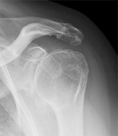
Caption: Pre-operative X-ray showing glenohumeral osteoarthritis - a narrowed joint space and a bone spur along the lower edge of the humeral head.
This is where nearly every shoulder replacement begins. The X-ray confirms arthritis: the cartilage space between ball and socket has narrowed, and a spur has formed along the bottom of the humeral head.
What the X-ray cannot tell me is the shape of the socket - how much bone has worn away, in which direction, and how far the whole surface has tilted. Those are three-dimensional questions, and this is a two-dimensional image.
Stage 2 - The CT that answers what the X-ray can’t
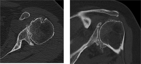
Caption: CT of the same shoulder, looking down through the joint. These slices show the pattern of glenoid wear and how far the socket has tilted backward.
Looking down through the joint, the erosion of the socket becomes visible - along with how far it has rotated backward, and how much bone remains to seat an implant against.
This scan is also the dataset the plan is built from. Everything that follows comes from these images.
Stage 3 - The plan
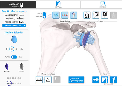
Caption: The pre-operative plan for this shoulder, built on a three-dimensional model generated from the CT scan above. Both components are positioned and sized before the day of surgery - here, a stemless humeral component and a glenoid component, shown seated on the patient’s own anatomy.
This is the plan. The model is the patient’s own bone, reconstructed from their CT, and both implants are positioned on it before the operation begins - size, position, version, inclination, and depth, with the software reporting how much of the component is backed by bone as the position is adjusted.
The measurements shown on the left are the software’s prediction of how the reconstructed joint will sit once the implants are in. Nothing here is estimated during surgery. The decisions are made in advance, on the patient’s real anatomy, where changing them costs nothing.
Stage 4 - The result
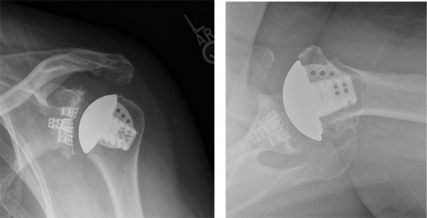
Caption: The same shoulder after surgery. AP and axillary X-rays showing the stemless humeral component and glenoid component sitting in the positions that were planned.
Vendor note: same asset also referenced on the Total Shoulder Replacement page.
These are the post-operative X-rays of the shoulder you have just followed from the first film. The implants sit where the plan said they would.
That is the proposition of navigation, stated as plainly as I can: the plan and the result are near identical. Not judged by eye at the end of a long case, but measured beforehand, executed, and then verified.
Case two: planning a stemmed replacement
Here is a different patient - this one receiving a stemmed implant rather than a stemless one - showing the two halves of the plan.
Planning the socket
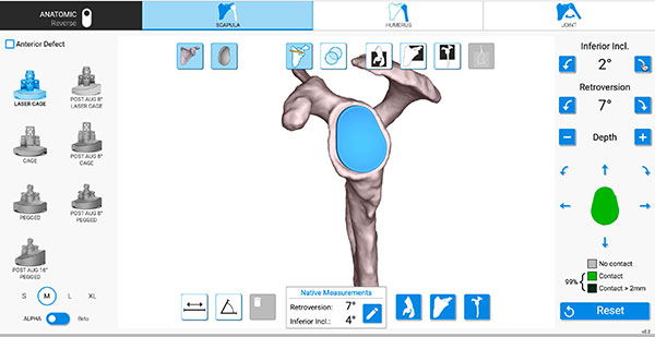
Caption: The glenoid component positioned on a three-dimensional model of the patient’s own scapula. The panel at the right shows the implant set to 2 degrees of inferior inclination against native measurements of 7 degrees retroversion and 4 degrees inclination - and reports 99% of the implant backed by bone.
The socket is planned first. Every measurement comes from the patient’s own CT rather than an estimate, and from there I work through implant type, size, position, version, inclination, and depth.
The percentage at the lower right is doing something specific and useful: it reports how much of the implant’s back surface is in contact with bone, and it updates as the position changes. Backing a component with bone rather than a gap is one of the things that gives it a chance of lasting. In this plan it reads 99%.
Planning the humeral side - and checking how the shoulder will move
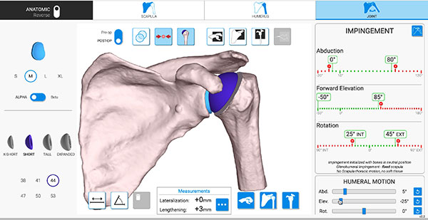
Caption: The same patient with both components in place and the joint assembled. The panel at the right reports the arc of motion the reconstruction allows before the components would begin to collide - abduction, forward elevation, and rotation, each measured.
The humeral side follows: stem length and size, the level and angle of the bone cut, retroversion, and inclination - each checked against the patient’s CT slices alongside the three-dimensional model.
Then comes the part most patients find surprising. With both components in place, the software assembles the joint and moves it, reporting the arc of motion the reconstruction will allow and the point at which the components would begin to collide. The range of motion is checked before the operation begins, not assessed afterwards.
Every one of these decisions is made in advance, on the patient’s real anatomy, where changing them costs nothing.
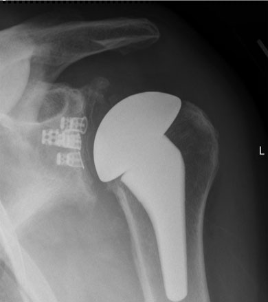
Caption: The result - anatomic total shoulder replacement with a stemmed humeral component. The stem extends down into the shaft of the humerus.
Vendor note: same asset also used on the Total Shoulder Replacement page.
What the evidence shows
In a published analysis of more than 16,000 navigated shoulder arthroplasty cases on this platform, 98% were completed as planned, with implant position verified to within approximately 2 millimeters and 2 degrees of the preoperative plan.
Additional findings from the published literature on GPS navigation in shoulder arthroplasty:
- Minimal deviation from planned glenoid version, inclination, and position across a large case series
- Improved range of motion and reduced postoperative complications compared with non-navigated groups in comparative studies
- An excellent safety profile - the most commonly reported navigation-specific complication, coracoid fracture at the tracker fixation site, occurred in 0.05% of cases
- Over 100,000 total joint cases performed on the GPS platform across shoulder, knee, and ankle applications
An honest framing of what this evidence does and does not establish. The data on accuracy is strong and consistent: navigation reliably improves how closely implant placement matches the surgical plan. The link from improved accuracy to improved long-term implant survival is biologically well-founded and supported by comparative studies, but proving it definitively requires the kind of fifteen- and twenty-year follow-up that a technology introduced in the last decade has not yet had time to generate. What can be said accurately today is that navigation makes the operation more precise and more reproducible, and that malposition is a known cause of failure. Any surgeon who tells you navigation guarantees a better result twenty years from now is overstating the evidence.
“Is this a robotic shoulder replacement?”
No - and the distinction is worth understanding, because many patients search for “robotic shoulder replacement” after hearing about robotic knee and hip surgery.
A robotic system physically constrains or executes part of the bone preparation. A navigation system does not touch the patient and does not move anything. It measures, guides, and verifies. Every cut, every reamer, every implant is placed by the surgeon’s hands.
Navigation is also the more established technology in this joint. The platform I use received FDA clearance for shoulder arthroplasty in May 2017, predating every robotic option available for the shoulder - so the accuracy data behind it reflects years of real-world use rather than early experience.
The case for guidance is arguably stronger in the shoulder than in the knee or hip, precisely because the glenoid is smaller, harder to see, and less forgiving of error.
I mention this because “surgeon-controlled” is not a marketing qualifier. It is an accurate description of how the technology works, and patients deserve to know what they’re actually getting.
Who benefits most?
Navigation is useful in essentially every shoulder replacement, but the benefit scales with complexity. It matters most in:
- Significant glenoid bone loss or severe retroversion - where the margin for error is smallest and freehand estimation is least reliable
- Reverse shoulder replacement - where baseplate position and screw trajectory into limited bone directly affect stability and fixation
- Cuff tear arthropathy - often accompanied by eroded, deformed glenoid anatomy
- Post-traumatic arthritis and deformity from prior fracture
- Revision shoulder arthroplasty, where anatomy is altered and bone stock compromised
- Younger patients, for whom the implant needs to last the longest and every degree of alignment compounds over more years
What this means for you as a patient
Practically speaking, choosing a navigated shoulder replacement means:
- You’ll have a CT scan before surgery rather than only X-rays.
- Your operation is planned in advance on a model of your own anatomy, not decided entirely in the moment.
- Surgery takes modestly longer - generally a matter of minutes, not hours.
- Your recovery is unchanged. Navigation affects how the implant is placed, not what you do afterward. Rehabilitation follows the same protocol as any anatomic or reverse
- Navigation is reported alongside the surgery using a specific add-on code. How that is handled varies between insurers - many bundle it into the payment for the operation itself. Our office verifies your benefits before surgery, so you will know what to expect rather than finding out afterwards.
Frequently Asked Questions
What is computer-navigated shoulder replacement?
It is shoulder replacement surgery performed using a computer navigation system that tracks surgical instruments in real time and compares their position against a three-dimensional plan built from the patient’s own preoperative CT scan. It allows the surgeon to verify that implant position, angle, and screw trajectory match the plan before anything is permanently fixed in place.
Is computer-navigated shoulder replacement the same as robotic surgery?
No. A robot physically performs or constrains part of the procedure. Navigation does neither - it is a measurement and verification system. The surgeon performs every step by hand, using the navigation display for real-time positional feedback. Navigation is also the longer-established technology in the shoulder: the platform used in this practice received FDA clearance for shoulder arthroplasty in May 2017, predating every robotic option available for this joint.
Does navigation make shoulder replacement safer?
The published safety profile is excellent, with the most common navigation-specific complication - coracoid fracture at the tracker site - reported in 0.05% of more than 16,000 cases. Navigation’s main contribution is accuracy: verified implant placement within roughly 2 mm and 2 degrees of plan, with 98% of cases completed as planned.
Does navigated surgery take longer?
Modestly. Registration and verification add a small amount of operative time, generally measured in minutes. Much of the planning work happens before the day of surgery, which can offset intraoperative decision-making time.
Does computer navigation cost more?
Navigation is reported alongside the surgery using a specific add-on billing code, and how insurers handle that code varies - many bundle it into the payment for the operation itself. Whether it affects your out-of-pocket cost depends on your particular plan, so it isn’t something to assume in either direction. Our office verifies your benefits before surgery so you know what to expect in advance. You will also need a preoperative CT scan, which is generally handled as part of standard surgical planning.
How do I know if my surgeon uses navigation?
Ask directly. Navigation is not yet standard in shoulder arthroplasty, and adoption varies considerably between surgeons and institutions. It is also worth asking how many navigated cases a surgeon has actually performed, since familiarity with the platform matters as much as access to it. By case volume, Dr. Ode ranks among the top 5% of surgeons worldwide using navigated shoulder arthroplasty.
Which patients benefit most from navigation?
Patients with significant glenoid bone loss or deformity, those undergoing reverse shoulder replacement, patients with cuff tear arthropathy or post-traumatic deformity, revision cases, and younger patients whose implants need to last the longest.
Interested in a navigated shoulder replacement?
Dr. Gabriella Ode is a fellowship-trained shoulder surgeon at the HSS Sports Medicine Institute and Associate Professor of Orthopaedic Surgery at Weill Cornell Medical College. She is nationally recognized for expertise in computer-navigated shoulder arthroplasty and performs both primary and revision shoulder replacement. Offices on the Upper East Side, the West Side, and in Brooklyn. Request an appointment or request a remote second opinion if you are outside the New York area.
Learn more about the technology: AdvitaGPS navigation system · GPS Shoulder application
Product-specific disclosure.
This article describes the AdvitaGPS navigation platform, manufactured by Advita Ortho, for whom Dr. Ode is a paid consultant. The clinical data cited here is drawn from peer-reviewed published literature rather than manufacturer materials, and the limitations of that evidence are stated in the text above.
Financial disclosure.
Dr. Ode is a paid consultant for Advita Ortho and ConMed, manufacturers of orthopaedic implants and surgical instrumentation. These relationships are reported publicly through the CMS Open Payments database. She receives no compensation for this website, and no compensation is tied to any individual patient’s treatment decision. Her complete financial disclosures are available here.
This article is for general education and is not a substitute for individual medical advice. Please consult a physician about your specific condition.